
What Is It?
Retinal vein occlusion (RVO) is a retinal vascular condition that occurs when blood flow through one of the veins draining the retina becomes blocked or significantly reduced.
The retina is the light-sensitive tissue at the back of the eye that converts light into visual signals. It requires healthy blood circulation to function properly.
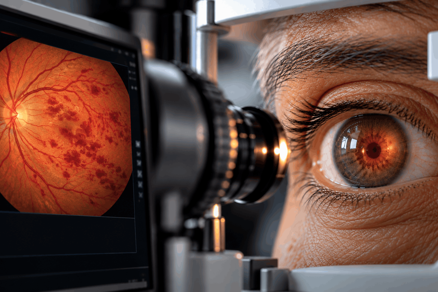
When a retinal vein becomes blocked, pressure increases within the affected blood vessels. This can cause blood and fluid to leak into the retina, leading to retinal hemorrhages, swelling, and impaired vision.
In some patients, reduced blood flow causes retinal ischemia, meaning that parts of the retina do not receive enough oxygen.
There are two main types of retinal vein occlusion:
1. Central Retinal Vein Occlusion (CRVO)
CRVO affects the main vein responsible for draining blood from the retina.
Because the central retinal vein drains most of the retinal circulation, a blockage can affect a large area of the retina and cause significant vision loss.
CRVO can be classified as ischemic or non-ischemic depending on the extent of impaired retinal blood flow.
2. Branch Retinal Vein Occlusion (BRVO)
BRVO occurs when one of the smaller branches of the retinal venous system becomes blocked.
It usually affects a particular region of the retina rather than the entire retinal circulation.
Visual symptoms depend on the location of the blockage and whether the central macula is affected.
A related form, hemiretinal vein occlusion, affects approximately one half of the retinal circulation.
One of the most important complications of RVO is macular edema, which occurs when fluid accumulates in the macula, the retinal area responsible for sharp central vision.
Severely reduced retinal blood flow may also stimulate the development of abnormal new blood vessels, which can cause further complications.
Retinal vein occlusion usually affects one eye, although both eyes can be affected.
Some patients recover useful vision, particularly with early treatment of complications, while others may experience persistent visual impairment.
Prompt diagnosis, individualized treatment, and ongoing retinal monitoring are essential for protecting vision.
Symptoms
Symptoms of retinal vein occlusion vary according to the location and severity of the blockage.
Some patients experience sudden visual changes, while others notice symptoms developing over hours or days.
Common symptoms include:
• Sudden blurred vision in one eye.
• Partial or substantial loss of vision.
• A dark or blurred area within the visual field.
• Distorted central vision.
• Difficulty reading or recognizing fine details.
• Reduced contrast or clarity of vision.
• Floaters, particularly when bleeding occurs inside the eye.
Mild retinal vein occlusion may occasionally produce few or no noticeable symptoms.
Symptoms are often painless, especially at the beginning of the condition.
In Central Retinal Vein Occlusion (CRVO), vision loss may be more widespread because a larger retinal area is affected.
In Branch Retinal Vein Occlusion (BRVO), symptoms may involve only part of the visual field or central vision, depending on the affected branch.
Advanced ischemic disease can occasionally lead to abnormal blood vessel growth and increased eye pressure, producing pain or redness.
IMPORTANT WARNING SIGNS
Any sudden loss or significant reduction of vision requires immediate medical assessment.
New severe eye pain, a red eye with reduced vision, or rapid worsening of vision also requires urgent ophthalmic evaluation.
Because sudden visual loss can have several serious causes, including retinal artery occlusion, patients should not attempt to diagnose retinal vein occlusion themselves or delay emergency care.
Causes
Retinal vein occlusion develops when normal blood drainage from the retina becomes obstructed.
The mechanisms can vary according to the type of occlusion and the patient's vascular health.
1. Compression of Retinal Veins
In branch retinal vein occlusion, a thickened or hardened retinal artery may compress a neighboring vein at a point where the two vessels cross.
This can disturb blood flow and contribute to clot formation.
2. Venous Thrombosis
A blood clot may form within a retinal vein, reducing or blocking the normal return of blood from the retina.
In central retinal vein occlusion, the blockage typically involves the main retinal vein near the optic nerve.
3. Vascular Changes
High blood pressure, diabetes, and atherosclerotic vascular disease can affect blood vessel health and contribute to retinal vascular obstruction.
4. Increased Eye Pressure and Glaucoma
Glaucoma and elevated intraocular pressure are associated with an increased risk of certain forms of retinal vein occlusion, particularly CRVO.
5. Less Common Underlying Conditions
In selected patients, especially younger individuals or those with unusual or recurrent disease, systemic inflammatory, hematological, or clotting disorders may contribute.
After an occlusion occurs, impaired venous drainage can cause:
• Retinal hemorrhages.
• Leakage of fluid into retinal tissue.
• Macular edema.
• Reduced retinal oxygen supply.
• Abnormal new blood vessel formation in ischemic cases.
The underlying cause is often multifactorial, and it may not be possible to identify one single trigger.
Risk Factors
Several conditions are associated with an increased risk of retinal vein occlusion.
Important risk factors include:
• Increasing age.
• High blood pressure.
• Diabetes mellitus.
• High cholesterol or abnormal blood lipid levels.
• Atherosclerosis and other cardiovascular risk factors.
• Glaucoma.
• Elevated intraocular pressure.
• Smoking.
• A previous retinal vein occlusion.
Less commonly, certain blood disorders, inflammatory diseases, or conditions affecting blood clotting may contribute, particularly in younger patients.
The pattern of risk factors differs between CRVO and BRVO.
Having one or more risk factors does not necessarily mean a retinal vein occlusion will occur.
Patients diagnosed with RVO should have an appropriate assessment of their general vascular health.
This may involve coordination between the ophthalmologist and the patient's primary care physician or other medical specialists.
Additional blood tests or investigations may be appropriate for selected patients, especially when the clinical presentation is unusual.
Diagnosis
Retinal vein occlusion is diagnosed through a detailed retinal examination, supported by specialized imaging when clinically indicated.
The ophthalmologist assesses the type of occlusion, the presence of macular edema, the extent of reduced retinal blood flow, and the risk of abnormal blood vessel growth.
1. Medical History and Visual Assessment
The doctor reviews the onset of symptoms and relevant medical conditions, including high blood pressure, diabetes, cardiovascular disease, and glaucoma.
Visual acuity testing measures the severity of visual impairment.
2. Dilated Fundus Examination
Dilating eye drops allow a detailed examination of the retina.
Typical findings may include retinal hemorrhages, dilated or tortuous veins, retinal swelling, and signs of impaired circulation.
The distribution of these findings helps distinguish CRVO from BRVO.
3. Optical Coherence Tomography (OCT)
OCT produces detailed cross-sectional images of the retina.
It is particularly useful for identifying macular edema and measuring retinal thickness.
Repeat OCT examinations help monitor treatment response.
4. Fundus Photography
Retinal photographs document hemorrhages, vascular changes, and other findings.
They can also help assess changes over time.
5. Fluorescein Angiography (FFA)
Fluorescein angiography uses an injected dye to examine retinal blood circulation.
It may help identify areas of capillary nonperfusion, vascular leakage, or abnormal blood vessel growth.
6. OCT Angiography (OCTA)
OCTA provides additional information about retinal microvascular circulation without an injected dye.
It may be useful in selected cases to assess changes in the retinal capillary networks.
7. Intraocular Pressure and Anterior Segment Assessment
Eye pressure is measured, and the front of the eye may be examined for abnormal new blood vessels, especially in ischemic CRVO.
Gonioscopy may be necessary in patients at risk of abnormal vessel growth in the drainage angle.
8. General Medical Assessment
Blood pressure measurement and assessment for diabetes, abnormal blood lipids, and other vascular risk factors may be recommended.
Additional laboratory investigations are selected according to the patient's age, medical history, and clinical findings.
Not every patient requires every test.
The ophthalmologist combines the examination and imaging results to determine the appropriate treatment and monitoring plan.
Treatment Options
Treatment of retinal vein occlusion focuses on preserving vision, treating retinal complications, and reducing the risk of further damage.
There is no established treatment that reliably reopens every blocked retinal vein.
The management plan depends on whether the patient has CRVO or BRVO, the degree of retinal ischemia, the presence of macular edema, and the response to treatment.
1. Intravitreal Anti-VEGF Injections
Anti-vascular endothelial growth factor (anti-VEGF) treatment is a principal first-line therapy for macular edema associated with retinal vein occlusion.
These medications are injected into the eye under sterile conditions after local anesthesia is applied.
Anti-VEGF therapy helps reduce retinal fluid leakage and macular swelling.
In many patients, treatment improves or stabilizes vision.
Repeated injections are often required, especially during the initial treatment period.
The ophthalmologist adjusts treatment intervals based on visual acuity, OCT findings, and disease activity.
2. Intravitreal Corticosteroid Treatment
Corticosteroid injections or implants may be considered for selected patients with macular edema.
These treatments can reduce retinal swelling but may increase intraocular pressure and accelerate cataract formation.
They are not suitable for every patient and require appropriate follow-up.
3. Retinal Laser Treatment
Laser photocoagulation may be used for certain complications of retinal vein occlusion.
Panretinal photocoagulation or targeted peripheral retinal laser may be required when abnormal new blood vessels develop as a result of retinal ischemia.
For selected cases of BRVO, focal or grid laser treatment may be considered for persistent macular edema.
Laser treatment is not automatically required for all patients with RVO.
4. Management of Abnormal Blood Vessel Growth
Severe retinal ischemia can stimulate neovascularization, including abnormal vessels on the iris, drainage angle, retina, or optic disc.
This may result in vitreous hemorrhage or neovascular glaucoma.
Treatment may involve retinal laser photocoagulation, anti-VEGF therapy, and additional procedures when necessary.
Close monitoring is especially important for patients with ischemic CRVO.
5. Observation and Regular Retinal Monitoring
Some patients with mild RVO and no vision-threatening macular edema or neovascular complications may not require immediate ocular injections or laser treatment.
However, regular retinal examinations are essential because complications can develop after the initial occlusion.
Follow-up may include:
• Visual acuity measurements.
• OCT imaging.
• Retinal examination.
• Assessment for abnormal blood vessels.
• Eye pressure monitoring when indicated.
6. Management of Systemic Risk Factors
Blood pressure, diabetes, abnormal cholesterol levels, and other vascular risk factors should be evaluated and managed in cooperation with the patient's medical team.
Controlling these conditions supports overall cardiovascular health and may help reduce future vascular risk.
Routine anticoagulant or antiplatelet medication is not an established treatment for restoring vision after an isolated retinal vein occlusion. Such medicines should only be used when indicated for the patient's general medical condition.
7. Management of Advanced Complications
In selected patients with complications such as persistent vitreous hemorrhage or other complex retinal abnormalities, additional retinal procedures or surgery may be considered.
Treatment and outcomes are individualized.
Some patients experience substantial visual improvement, while others have persistent impairment due to macular ischemia, chronic edema, or irreversible retinal damage.
Early specialist care and appropriate long-term follow-up are important for protecting remaining vision.
Prevention & Care
Not all retinal vein occlusions can be prevented, but careful management of vascular and eye health may help reduce certain risks.
Recommended measures include:
• Monitor and manage blood pressure.
• Maintain appropriate diabetes control.
• Manage abnormal cholesterol and other cardiovascular risk factors.
• Avoid smoking.
• Maintain regular physical activity and healthy lifestyle habits.
• Attend recommended eye examinations, especially if you have glaucoma.
• Follow the retinal specialist's treatment plan.
• Attend scheduled follow-up visits even if vision improves.
• Report any new or worsening visual symptoms promptly.
• Discuss your general vascular health with your primary care physician.
Why Is Follow-Up Important?
Retinal vein occlusion can cause delayed complications, including recurrent or persistent macular edema and abnormal blood vessel growth.
Even when vision improves, ongoing retinal monitoring may be necessary.
Patients with ischemic CRVO may require particularly close follow-up because abnormal blood vessels can develop in the iris or drainage angle and lead to neovascular glaucoma.
When Should You See an Eye Doctor?
Seek immediate medical assessment for:
• Sudden or unexplained loss of vision.
• A rapid reduction in vision in one eye.
• A new dark area or major visual field defect.
• Severe eye pain or a painful red eye associated with reduced vision.
Do not wait for a routine appointment when sudden vision loss occurs.
General Health Considerations
A retinal vein occlusion may occur alongside important cardiovascular risk factors.
Patients should follow medical advice regarding blood pressure, diabetes, cholesterol, and any relevant systemic conditions.
Do not start or stop blood thinners or other prescribed medications without guidance from the treating physician.
Early detection of complications and consistent follow-up help protect long-term visual function.
Frequently Asked Questions
What is retinal vein occlusion?
What is the difference between CRVO and BRVO?
What causes retinal vein occlusion?
Is retinal vein occlusion painful?
Can retinal vein occlusion cause sudden vision loss?
Can retinal vein occlusion cause blindness?
Can retinal vein occlusion be cured?
What is macular edema?
Are eye injections necessary for every patient?
How many anti-VEGF injections will I need?
Is laser treatment used for retinal vein occlusion?
Can vision improve after treatment?
Can retinal vein occlusion happen again?
Is retinal vein occlusion related to high blood pressure?
Do I need blood-thinning medication?
How is retinal vein occlusion diagnosed?
Why are follow-up appointments important?
When should I seek urgent medical care?
Our team can help you with an individual medical assessment.