
What Is It?
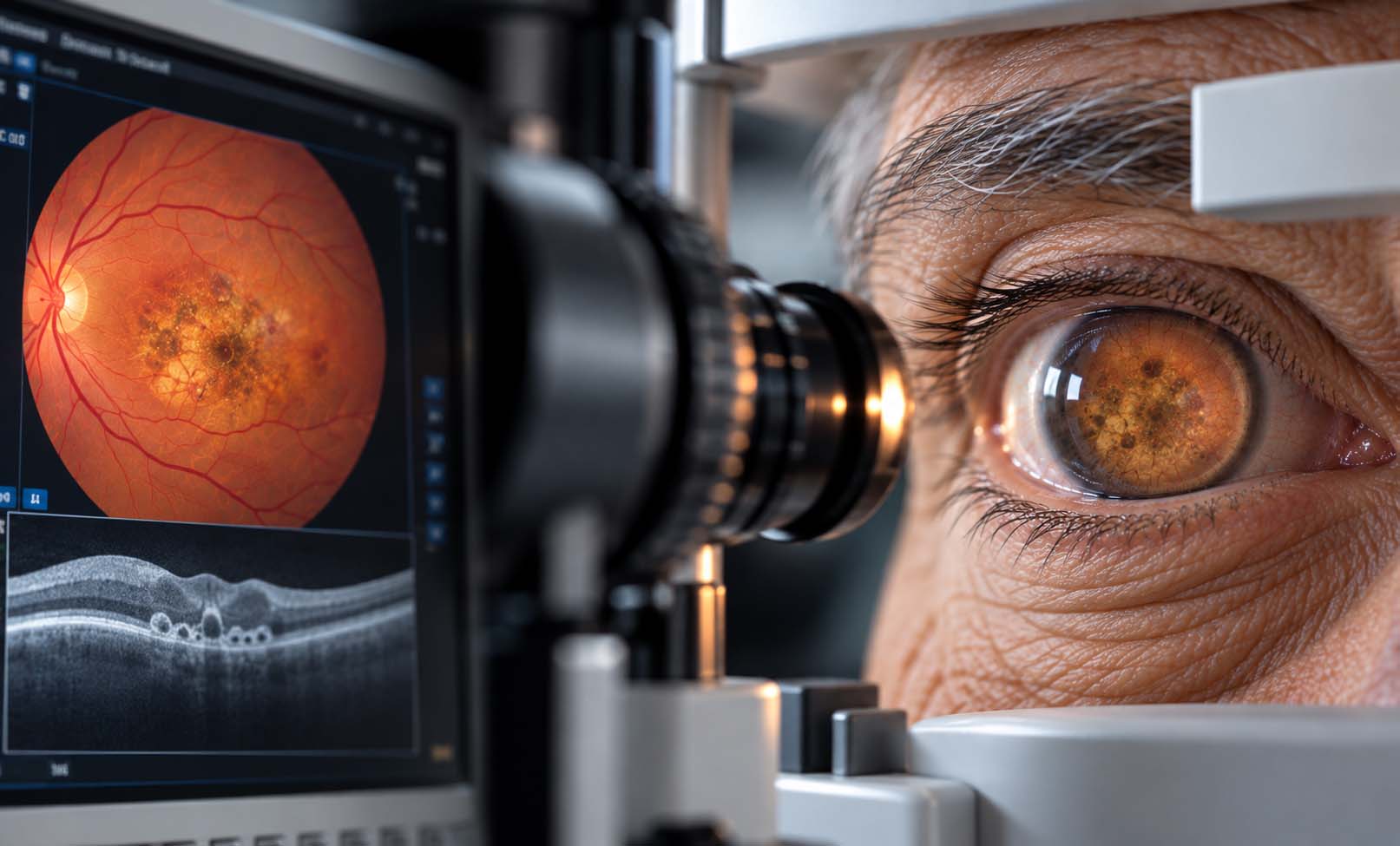
Age-related macular degeneration (AMD) is a condition that affects the macula, the central part of the retina responsible for detailed, straight-ahead vision.
The macula allows us to perform activities such as reading, driving, recognizing faces, and seeing fine details.
AMD mainly affects adults over the age of 50. It may develop in one or both eyes, and its progression varies between individuals.
There are two main forms of AMD:
Dry AMD (Non-Neovascular AMD)
Dry AMD is the more common form. It is associated with changes in the macula, including the accumulation of small deposits called drusen and damage to retinal cells.
Dry AMD is classified into early, intermediate, and late stages. Early disease may cause no noticeable symptoms, while advanced dry AMD can lead to geographic atrophy, in which areas of retinal tissue gradually deteriorate.
Wet AMD (Neovascular AMD)
Wet AMD occurs when abnormal blood vessels grow beneath the retina or retinal pigment epithelium. These fragile vessels may leak fluid or blood, damaging the macula.
Wet AMD can cause relatively rapid central vision loss and requires prompt specialist assessment and treatment.
AMD primarily affects central vision. Peripheral vision is often preserved, although advanced disease may substantially interfere with everyday activities.
There is no complete cure for AMD, but early detection, appropriate treatment, and ongoing monitoring can help preserve vision and support quality of life.
Symptoms
Symptoms of age-related macular degeneration depend on its stage and whether the condition is dry or wet.
Early AMD may cause no noticeable symptoms.
As the disease progresses, symptoms may include:
• Blurred or reduced central vision.
• Difficulty reading small print or recognizing faces.
• Straight lines appearing wavy, bent, or distorted.
• A dark, blurry, or missing area in the center of vision.
• Difficulty seeing fine details.
• Increased difficulty seeing in dim lighting.
• Colors appearing less bright or vivid.
• Needing brighter light when reading.
Dry AMD often develops gradually, although progression varies.
Wet AMD may cause new distortion or a more rapid decline in central vision.
Important Warning Signs
Sudden or recent distortion of straight lines, a new central dark spot, or a rapid change in central vision requires prompt assessment by an ophthalmologist or retina specialist.
Changes may be less noticeable when one eye is affected, because the other eye can compensate. Each eye should therefore be assessed separately.
Causes
The exact cause of age-related macular degeneration is not fully understood.
AMD develops through a combination of age-related retinal changes, genetic susceptibility, and environmental factors.
Over time, the macula may undergo changes affecting the retinal pigment epithelium, photoreceptor cells, and supporting tissues.
In dry AMD, characteristic changes may include:
• Accumulation of drusen beneath the retina.
• Dysfunction of retinal pigment epithelial cells.
• Gradual damage to light-sensitive retinal cells.
• Development of geographic atrophy in advanced disease.
In wet AMD, abnormal blood vessels develop beneath the retina or retinal pigment epithelium. These vessels may leak fluid or blood, leading to swelling, distortion, and damage to central vision.
Dry AMD can progress to late dry AMD or develop a wet component.
Not everyone with early AMD develops advanced disease. The rate and pattern of progression differ from patient to patient.
Risk Factors
Several factors are associated with an increased risk of age-related macular degeneration.
Established and important risk factors include:
• Increasing age, particularly after age 50.
• Family history of AMD.
• Smoking.
• Genetic susceptibility.
• Previous AMD in the other eye.
Other factors associated with AMD risk or progression may include:
• High blood pressure and cardiovascular risk factors.
• Obesity.
• Limited physical activity.
• Diets low in vegetables and other nutrient-rich foods.
Having risk factors does not mean a person will necessarily develop AMD.
Smoking is an important modifiable risk factor. People with a family history of AMD should discuss their individual need for regular retinal examinations with an eye care professional.
Diagnosis
Age-related macular degeneration is diagnosed through a comprehensive retinal examination supported by imaging when appropriate.
The ophthalmologist may use the following assessments:
1. Medical History and Visual Assessment
The doctor reviews symptoms, family history, smoking history, general health, and changes in everyday visual activities.
Visual acuity testing measures the clarity of central vision.
2. Dilated Fundus Examination
Pupil-dilating eye drops allow detailed examination of the macula and surrounding retina.
The ophthalmologist looks for drusen, pigment changes, retinal atrophy, fluid, bleeding, or other abnormalities.
3. Optical Coherence Tomography (OCT)
OCT is a non-invasive imaging test that produces detailed cross-sectional images of the retina.
It helps identify retinal fluid, macular structural changes, atrophy, and signs of neovascular AMD.
OCT is particularly important in diagnosing and monitoring wet AMD.
4. OCT Angiography (OCTA)
OCTA may help visualize abnormal retinal or choroidal blood vessel networks without an injected dye.
It is used selectively according to the clinical findings.
5. Fluorescein Angiography (FFA)
In certain cases, fluorescein angiography helps detect abnormal leaking blood vessels and clarify the diagnosis of wet AMD.
6. Amsler Grid Assessment
An Amsler grid can help identify distortion or missing areas of central vision.
It may also be used for home monitoring, but it does not replace professional eye examinations or retinal imaging.
Not every patient requires every investigation. The ophthalmologist selects the appropriate tests based on symptoms and examination findings.
Treatment Options
Treatment for age-related macular degeneration depends on the type and stage of the disease.
The primary goals are to preserve remaining vision, reduce the risk of progression, and support everyday visual function.
1. Management of Early Dry AMD
Early dry AMD usually does not require an eye injection or surgical procedure.
Management includes regular eye examinations, smoking cessation, healthy lifestyle measures, and monitoring for visual changes.
Patients should report new distortion or deterioration in central vision promptly.
2. AREDS2 Nutritional Supplements
For selected patients with intermediate AMD or advanced AMD in one eye, the AREDS2 vitamin and mineral formulation may reduce the risk of progression to advanced AMD.
These supplements are not recommended automatically for everyone with AMD and do not cure existing retinal damage.
An ophthalmologist should assess whether the patient meets the appropriate criteria and review possible medication interactions or other health considerations.
3. Intravitreal Anti-VEGF Injections for Wet AMD
Anti-vascular endothelial growth factor (anti-VEGF) therapy is a principal treatment for active wet AMD.
These medications are injected into the eye under sterile conditions after local anesthetic is applied.
Anti-VEGF treatment helps control abnormal blood vessel growth and leakage.
It can stabilize vision and may improve vision in some patients, particularly when treatment begins promptly.
Patients usually require repeated injections and regular OCT monitoring. The number and timing of injections depend on disease activity and response.
4. Treatment of Geographic Atrophy
Geographic atrophy is an advanced form of dry AMD that causes progressive loss of retinal tissue.
Complement-inhibitor intravitreal therapies have become available in certain countries for selected patients with geographic atrophy.
These treatments may slow the enlargement of atrophic lesions but do not restore retinal cells that have already been lost.
Their suitability, safety considerations, regulatory approval, and local availability must be discussed with a retina specialist.
5. Other Procedures in Selected Cases
Photodynamic therapy or other specialist procedures may be considered in selected neovascular conditions, depending on the diagnosis and clinical findings.
They are not routine treatments for every patient with AMD.
6. Low Vision Rehabilitation
Patients with persistent central visual impairment may benefit from:
• Magnifying devices and electronic reading aids.
• Improved lighting and contrast.
• Low vision assessment and rehabilitation.
• Accessibility features on phones, tablets, and computers.
• Practical support for reading and daily activities.
AMD management should be individualized by an ophthalmologist or retina specialist. Early recognition of wet AMD and consistent follow-up are especially important.
Prevention & Care
Age-related macular degeneration cannot always be prevented, but several measures can help support retinal health and reduce certain risks.
Recommended eye care measures include:
• Avoid smoking and seek support to quit if needed.
• Attend eye examinations according to your ophthalmologist's recommendations.
• Follow a balanced diet that includes leafy green vegetables and other nutrient-rich foods.
• Maintain regular physical activity and a healthy lifestyle.
• Manage blood pressure and other cardiovascular risk factors.
• Take AREDS2 supplements only when recommended for your stage of AMD.
• Follow prescribed treatment and monitoring schedules.
• Use appropriate visual aids when necessary.
Monitoring Vision at Home
Patients with AMD may be advised to monitor each eye separately for changes in central vision.
An Amsler grid is one possible monitoring tool.
When using an Amsler grid, patients should wear their usual reading glasses if required, cover one eye, look at the center point, and notice whether lines appear distorted or areas seem missing.
A normal Amsler grid result does not rule out worsening AMD.
When Should You See an Eye Doctor?
Arrange prompt ophthalmic assessment if you notice:
• New distortion of straight lines.
• A new blurred, dark, or missing central area.
• Sudden or rapid deterioration in central vision.
• A noticeable change in vision in either eye.
Early recognition and treatment of wet AMD can help reduce the risk of irreversible visual damage.
Frequently Asked Questions
What is age-related macular degeneration?
What is the difference between dry and wet AMD?
Can macular degeneration cause blindness?
Is macular degeneration painful?
Can AMD be cured?
Can dry AMD become wet AMD?
Can wet AMD be treated with eye injections?
Are vitamin supplements useful for every patient with AMD?
What is geographic atrophy?
Can lost vision from AMD return?
How is AMD diagnosed?
What should I do if straight lines suddenly look wavy?
Can AMD affect both eyes?
Does AMD always require surgery?
Our team can help you with an individual medical assessment.