
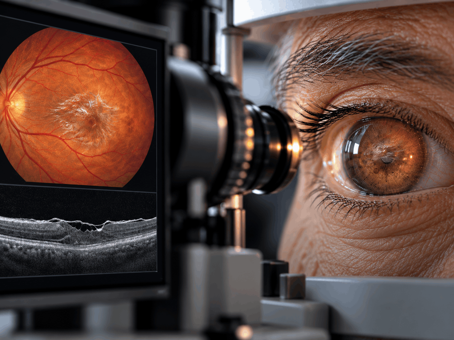
What Is It?
An epiretinal membrane (ERM) is a thin, semi-transparent layer of fibrous and cellular tissue that forms on the inner surface of the retina. When it develops over the macula, the central part of the retina responsible for sharp, detailed vision, it is commonly called a macular pucker.
The retina is the light-sensitive tissue lining the back of the eye. Its central region, the macula, allows us to read, recognize faces, distinguish fine details, and perform activities that require precise central vision.
An epiretinal membrane may initially be very thin and cause little or no disturbance. Over time, however, the membrane can contract and exert traction on the retinal surface. This pulling effect can create wrinkles or structural changes in the macula, interfering with the normal processing of visual images.
Patients may notice that straight lines appear bent or wavy, objects seem distorted, or small details become harder to see. These symptoms are described as metamorphopsia and may occur alongside reduced central visual clarity.
Epiretinal membranes commonly affect one eye, although both eyes can be involved. In some patients, the condition is detected incidentally during a routine retinal examination.
The condition is usually slowly progressive or relatively stable. Many people maintain functional vision without requiring surgery.
An epiretinal membrane is different from a full-thickness macular hole. However, it may be associated with changes in macular structure, including a macular pseudohole or, less commonly, other traction-related abnormalities.
Treatment depends on the severity of the membrane, the degree of visual distortion, changes observed on retinal imaging, and the impact on the patient's everyday activities.
Symptoms
Symptoms of an epiretinal membrane vary according to the thickness of the membrane, the amount of traction it creates, and the location of the affected retinal tissue.
Many patients with a mild epiretinal membrane have no noticeable symptoms. When symptoms occur, they commonly include:
• Blurred or reduced central vision, especially when looking at fine details.
• Distorted vision (metamorphopsia), in which straight lines appear wavy, curved, or irregular.
• Difficulty reading, particularly when letters appear crowded, displaced, or unclear.
• Difficulty recognizing faces or seeing fine details at a distance.
• Objects appearing smaller than normal in the affected eye (micropsia).
• A difference in image size or shape between the two eyes.
• Double or overlapping images perceived by one eye in some cases.
• Reduced visual comfort when performing detailed tasks.
Symptoms usually develop gradually and may remain mild for an extended period.
Because the unaffected eye can compensate, some patients do not recognize the problem until they cover one eye during reading or undergo an eye examination.
An epiretinal membrane mainly affects central vision rather than peripheral vision. It does not usually cause pain, redness, or total blindness.
Any sudden loss of vision, new flashes of light, a sudden increase in floaters, or the appearance of a dark curtain across the visual field requires urgent ophthalmic assessment, as these symptoms may indicate another retinal emergency.
Causes
The exact cause of an epiretinal membrane is not always identifiable. Most cases occur without a specific underlying retinal disease and are described as idiopathic epiretinal membranes.
A common association is the natural aging process of the vitreous, the transparent gel that fills the inside of the eye.
As people age, the vitreous gradually changes in structure and may separate from the retinal surface. This process is called posterior vitreous detachment (PVD).
In some eyes, microscopic changes at the vitreoretinal interface allow cells to proliferate on the retinal surface. These cells can form a thin fibrous membrane.
The membrane may subsequently contract, producing traction and wrinkling of the macula.
Epiretinal membranes can also develop secondary to other ocular conditions or events, including:
• Retinal tears or retinal detachment.
• Previous retinal or intraocular surgery.
• Retinal vein occlusion.
• Diabetic retinopathy and other retinal vascular disorders.
• Uveitis or inflammation inside the eye.
• Eye trauma.
• Certain retinal laser procedures or other conditions affecting the retinal surface.
The membrane is not a tumor or an infectious growth. It is a form of abnormal cellular and fibrous tissue on the retinal surface.
In many cases, the underlying mechanism is related to changes in the vitreoretinal interface rather than a single preventable cause.
Risk Factors
Several factors are associated with an increased likelihood of developing an epiretinal membrane.
Increasing Age
Epiretinal membranes are more frequently identified in people over 50 years of age, particularly in older adults.
Posterior Vitreous Detachment
Separation of the vitreous gel from the retina is a common age-related event and is strongly associated with the development of some epiretinal membranes.
Previous Retinal Tear or Detachment
Retinal injuries and the healing processes that follow can contribute to membrane formation.
Previous Eye Surgery
Certain intraocular procedures, including retinal surgery, may be associated with subsequent epiretinal membrane development.
Diabetic Retinopathy
Diabetes-related retinal vascular disease can increase the risk of secondary membrane formation.
Retinal Vein Occlusion
Vascular blockage and its associated retinal changes may contribute to fibrous tissue development on the retinal surface.
Ocular Inflammation
Conditions such as uveitis can trigger inflammatory and cellular responses that increase the likelihood of membrane formation.
Eye Trauma
Previous injury to the eye can produce retinal or vitreoretinal changes associated with secondary epiretinal membranes.
Epiretinal Membrane in the Other Eye
Patients with an ERM in one eye have an increased likelihood of developing a membrane in the fellow eye.
Having one or more risk factors does not mean that a person will necessarily develop an epiretinal membrane. The condition can also occur in individuals without any known previous ocular disease.
Diagnosis
An epiretinal membrane is diagnosed through a comprehensive ophthalmic examination, with particular attention to the retina and macula.
The assessment generally includes the following:
1. Medical and Visual History
The ophthalmologist asks about blurred or distorted vision, difficulties with reading, the duration of symptoms, previous eye surgery, retinal disease, diabetes, inflammation, or eye trauma.
2. Best-Corrected Visual Acuity Testing
Vision is measured with appropriate optical correction to assess the effect of the membrane on central visual clarity.
3. Dilated Fundus Examination
Pupil-dilating drops allow the ophthalmologist to examine the macula and the surrounding retina. A reflective or wrinkled appearance on the macular surface may suggest an epiretinal membrane.
4. Optical Coherence Tomography (OCT)
OCT is a central investigation in the diagnosis and monitoring of epiretinal membranes. This non-invasive imaging technique produces high-resolution cross-sectional images of the retinal layers.
OCT can reveal:
• The presence and extent of the membrane.
• Distortion or thickening of the macula.
• Changes in the normal foveal contour.
• Retinal traction or associated fluid.
• The presence of an associated macular pseudohole or other structural abnormalities.
5. Amsler Grid Testing
An Amsler grid may be used to assess distortion of central vision. Patients may notice that straight lines appear wavy, broken, or displaced.
6. Additional Retinal Assessment
Depending on the clinical findings, the ophthalmologist may perform further investigations to identify associated retinal vascular disease, inflammation, or other causes of visual impairment.
Repeated OCT examinations are particularly useful for monitoring changes over time and helping determine whether observation or surgical intervention is appropriate.
Treatment Options
The management of an epiretinal membrane depends on the severity of visual symptoms, the degree of macular distortion, OCT findings, and the patient's individual visual needs.
Not every epiretinal membrane requires treatment. Many remain stable and can be managed successfully through observation.
1. Observation and Regular Monitoring
Mild epiretinal membranes that cause little or no visual disturbance are commonly observed rather than treated surgically.
Regular ophthalmic examinations and OCT imaging may be recommended to monitor retinal thickness, membrane traction, and any changes in visual function.
The frequency of follow-up is determined by the ophthalmologist according to the condition's severity and progression.
2. Visual Assessment and Optical Correction
An updated glasses prescription may improve vision if a refractive error is also present.
However, glasses and contact lenses cannot remove the epiretinal membrane or reverse structural distortion of the macula.
Patients with significant visual distortion may continue to experience symptoms despite wearing the correct prescription.
3. Vitrectomy with Epiretinal Membrane Peeling
When the membrane causes significant visual distortion, reduced visual acuity, or difficulty with daily activities, vitreoretinal surgery may be considered.
The standard surgical approach is pars plana vitrectomy combined with removal of the epiretinal membrane.
During surgery, the retinal surgeon removes the vitreous gel to gain access to the retinal surface. Delicate microsurgical instruments are then used to carefully peel the membrane away from the macula.
In selected cases, the internal limiting membrane (ILM), a very thin layer on the retinal surface, may also be peeled to reduce the likelihood of recurrence.
Removing the membrane relieves its pulling effect and allows the macula to gradually relax toward a more natural configuration.
4. Recovery After Surgery
Visual recovery is usually gradual rather than immediate.
Many patients experience improvement in visual distortion and central visual clarity over the following months. In some cases, improvement may continue for six to twelve months or longer.
The amount of visual recovery varies depending on factors such as:
• The duration of the membrane.
• The severity of preoperative visual distortion.
• The condition of the retinal layers on OCT.
• The presence of associated retinal diseases.
• The patient's preoperative visual acuity.
Surgery does not guarantee completely normal vision, and some distortion may persist.
5. Surgical Risks and Considerations
As with other intraocular procedures, vitrectomy carries potential risks, including retinal tears or detachment, infection, bleeding, and changes in eye pressure.
Cataract progression is common after vitrectomy in patients who still have their natural crystalline lens.
The ophthalmologist discusses the expected benefits, limitations, and possible complications before recommending surgery.
There are currently no established eye drops, oral medications, supplements, or eye exercises that can reliably dissolve or remove an epiretinal membrane.
The decision to operate is individualized and generally based on the degree to which the condition affects useful vision and quality of life.
Prevention & Care
There is no established method that reliably prevents the formation of an epiretinal membrane, particularly when it develops as part of age-related vitreoretinal changes.
Nevertheless, regular eye care and appropriate management of associated conditions can help preserve retinal health and detect problems early.
Recommended measures include:
• Attend regular comprehensive eye examinations, particularly after age 50.
• Follow the ophthalmologist's recommended monitoring schedule if an epiretinal membrane has already been diagnosed.
• Undergo OCT imaging when advised to monitor changes in macular structure.
• Monitor central vision separately in each eye to recognize any new distortion.
• Use an Amsler grid if recommended by the ophthalmologist.
• Report increasing difficulty with reading, facial recognition, or other central-vision tasks.
• Manage diabetes and other systemic conditions that may affect retinal health.
• Seek appropriate treatment for uveitis, retinal vascular disease, or other existing eye conditions.
• Follow postoperative instructions carefully if vitreoretinal surgery has been performed.
• Avoid missing scheduled follow-up appointments after retinal surgery.
An epiretinal membrane may remain stable for years, and many patients do not require surgical treatment.
However, gradual worsening of visual distortion or a meaningful decline in vision should prompt reassessment by a retinal specialist.
Sudden vision loss, a curtain-like shadow, new flashes, or a sudden shower of floaters are not typical of a slowly progressing epiretinal membrane and require urgent evaluation to exclude retinal tears or detachment.
Frequently Asked Questions
What is an epiretinal membrane?
Is an epiretinal membrane the same as a macular pucker?
What are the main symptoms of an epiretinal membrane?
Can an epiretinal membrane cause blindness?
Does every epiretinal membrane need surgery?
How is an epiretinal membrane diagnosed?
Can glasses correct vision affected by an epiretinal membrane?
Can eye drops dissolve an epiretinal membrane?
What surgery is used to treat an epiretinal membrane?
Is epiretinal membrane surgery painful?
How long does visual recovery take after surgery?
Will vision return completely to normal after surgery?
Can an epiretinal membrane return after surgery?
What is the difference between an epiretinal membrane and a macular hole?
When should I see a retinal specialist?
Our team can help you with an individual medical assessment.