
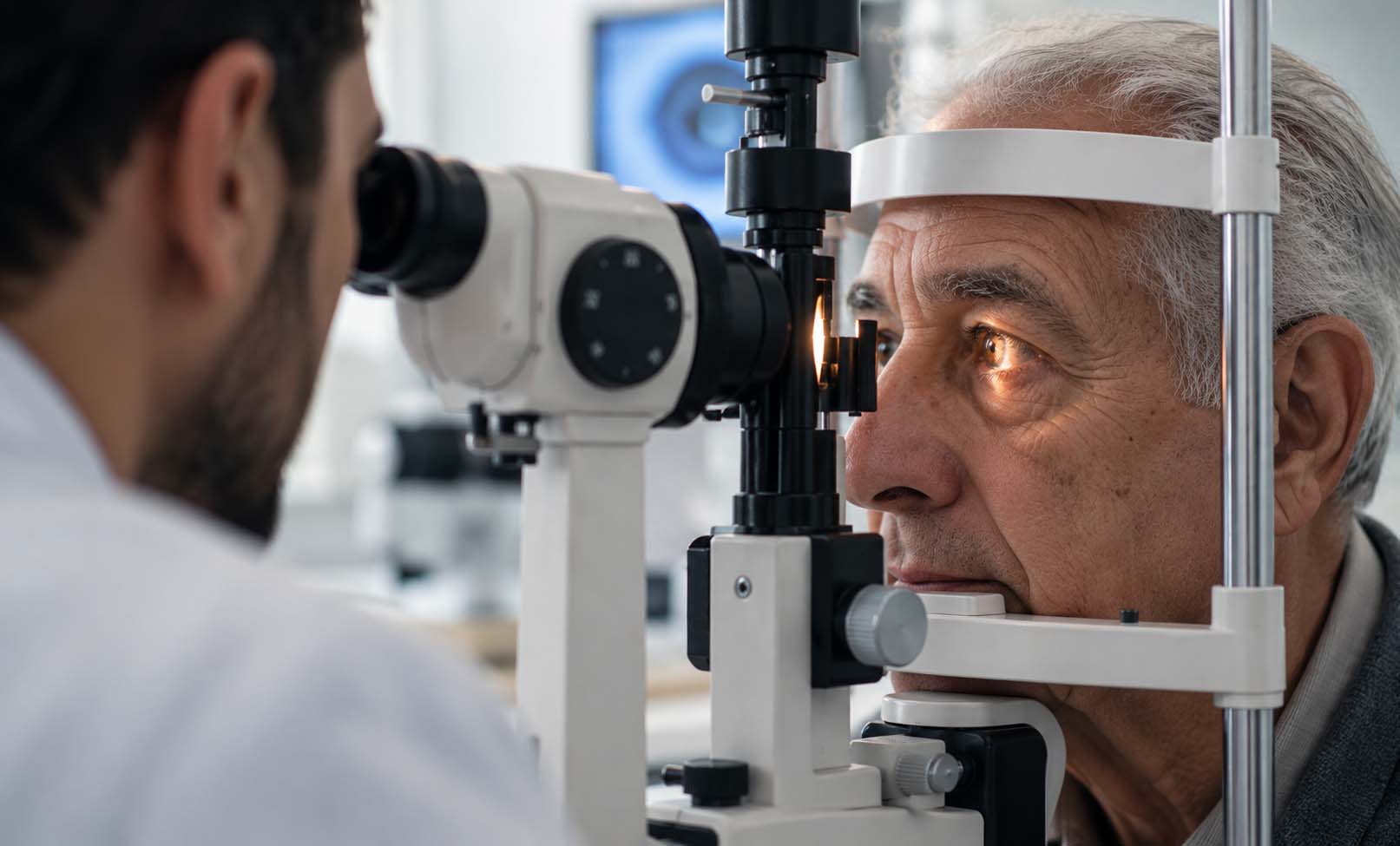
What Is It?
Glaucoma is a group of eye conditions that progressively damage the optic nerve, which carries visual information from the eye to the brain.
The damage is frequently associated with increased intraocular pressure (IOP), but glaucoma can also occur when eye pressure is within the normal range.
In many patients, glaucoma develops gradually, initially affecting peripheral (side) vision. Because these changes may go unnoticed, significant optic nerve damage can occur before symptoms become apparent.
The most common form is primary open-angle glaucoma. Other forms include angle-closure glaucoma, normal-tension glaucoma, secondary glaucoma, and childhood glaucoma.
Vision already lost from glaucoma generally cannot be restored. However, early diagnosis, appropriate treatment, and regular monitoring can help slow or prevent further vision loss.
Symptoms
Symptoms
Symptoms depend on the type and stage of glaucoma.
Open-Angle Glaucoma
The most common form typically progresses slowly and without pain.
Possible signs as the disease advances include:
Gradual loss of peripheral (side) vision.
Blind spots within the visual field.
Difficulty noticing objects to the side.
Reduced ability to navigate unfamiliar environments.
Severe visual impairment in advanced stages.
Many patients experience no noticeable symptoms during the early stages.
Acute Angle-Closure Glaucoma
A sudden rise in eye pressure can cause:
Severe eye pain.
Redness of the eye.
Sudden blurred or reduced vision.
Halos around lights.
Headache.
Nausea or vomiting.
Acute angle-closure glaucoma is a medical emergency. Anyone experiencing these symptoms should seek immediate emergency eye care.
Causes
Glaucoma develops when the optic nerve becomes damaged. The exact mechanisms vary according to the type of glaucoma.
In many forms, elevated intraocular pressure contributes to optic nerve damage.
The eye continuously produces a clear fluid called aqueous humor, which normally drains through structures in the front of the eye. When drainage becomes impaired, pressure inside the eye may rise.
In open-angle glaucoma, fluid drainage through the trabecular meshwork becomes less efficient.
In angle-closure glaucoma, the drainage angle between the iris and cornea becomes narrowed or blocked.
Some patients develop glaucomatous optic nerve damage despite eye pressure measurements within the statistically normal range. This is known as normal-tension glaucoma.
Glaucoma may also occur secondary to eye injuries, inflammation, certain medications, or other eye disorders.
Risk Factors
Factors associated with an increased risk of glaucoma include:
Increasing age.
A family history of glaucoma, especially in a first-degree relative.
Elevated intraocular pressure.
Certain ethnic backgrounds associated with higher risks of specific glaucoma types.
Thin central corneal thickness.
Significant nearsightedness (myopia), particularly in relation to open-angle glaucoma.
Certain anatomical features of the eye, including a narrow drainage angle.
Previous eye injury or inflammation.
Prolonged corticosteroid use in susceptible individuals.
Some forms of glaucoma may also occur in childhood.
Having a risk factor does not necessarily mean that glaucoma will develop, but it may indicate a need for more frequent eye examinations.
Diagnosis
Glaucoma is diagnosed through a comprehensive eye examination that evaluates the optic nerve, eye pressure, and visual function.
Depending on the patient's clinical findings, investigations may include:
Intraocular Pressure Measurement (Tonometry): Measures the pressure inside the eye.
Optic Nerve Examination: Evaluates the appearance and health of the optic nerve.
Optical Coherence Tomography (OCT): Provides detailed imaging of the optic nerve head and retinal nerve fiber layers to assess structural changes.
Visual Field Testing (Perimetry): Detects areas of peripheral or central vision loss related to glaucoma.
Gonioscopy: Examines the drainage angle of the eye to help identify open-angle or angle-closure mechanisms.
Pachymetry: Measures central corneal thickness, which can affect eye pressure interpretation and risk assessment.
Dilated Fundus Examination: Allows a detailed examination of the optic nerve and retina.
No single test can reliably diagnose every case of glaucoma. The ophthalmologist interprets the findings together and may repeat tests over time to detect progression.
Treatment Options
The primary goal of glaucoma treatment is to reduce the risk of further optic nerve damage and preserve the patient's remaining vision.
Lowering intraocular pressure is currently the main established treatment strategy, including for many patients with normal-tension glaucoma.
Treatment is individualized according to the glaucoma type, severity, progression risk, eye pressure, general health, and patient preferences.
1. Medicated Eye Drops
Prescription eye drops are commonly used to lower intraocular pressure.
Different medications work by reducing fluid production inside the eye, improving fluid drainage, or both.
Patients should use prescribed drops consistently and inform their ophthalmologist about side effects or difficulties administering treatment.
2. Laser Treatment
Laser procedures can help reduce eye pressure in selected patients.
Selective Laser Trabeculoplasty (SLT) is an established treatment for open-angle glaucoma and may be considered as an initial treatment for appropriate patients.
Laser peripheral iridotomy may be recommended for certain patients with angle-closure disease, depending on their clinical condition.
Additional treatment or repeat procedures may be necessary over time.
3. Glaucoma Surgery
Surgery may be recommended when the disease is advanced, pressure remains inadequately controlled, or further progression occurs despite other treatment.
Surgical options may include:
Trabeculectomy.
Glaucoma drainage implants.
Minimally invasive glaucoma surgery (MIGS) in selected patients.
Other pressure-lowering procedures, depending on the glaucoma type and severity.
The ophthalmologist will explain which treatment is suitable and discuss its expected benefits, risks, and recovery requirements.
4. Long-Term Monitoring
Glaucoma is generally a chronic condition requiring ongoing follow-up.
Regular pressure measurements, optic nerve imaging, and visual field testing help assess whether treatment is adequately controlling the disease.
Treatment can protect remaining vision, but established glaucomatous vision loss is generally irreversible.
Prevention & Care
There is no guaranteed way to prevent glaucoma. However, early detection and consistent treatment can substantially reduce the risk of progressive vision loss.
Recommended eye care measures include:
Attend comprehensive eye examinations at intervals appropriate to your individual risk.
Inform your ophthalmologist if a close family member has glaucoma.
Follow the prescribed treatment plan and attend scheduled follow-up appointments.
Do not stop glaucoma medications without consulting your treating ophthalmologist.
Discuss corticosteroid use with your healthcare professional, especially if treatment is prolonged.
Maintain good control of existing medical conditions as part of general health care.
Seek urgent medical attention for severe eye pain, sudden blurred vision, red eye, or associated nausea and vomiting.
Because early glaucoma often causes no noticeable symptoms, routine eye examinations are particularly important for people at increased risk.
Frequently Asked Questions
What is glaucoma?
Is glaucoma the same as high eye pressure?
Can glaucoma be cured?
Does glaucoma always cause symptoms?
Can glaucoma cause blindness?
Is glaucoma painful?
How is glaucoma diagnosed?
Can glaucoma be treated without surgery?
Will I need glaucoma treatment for life?
Can lost vision from glaucoma be restored?
Our team can help you with an individual medical assessment.