
What Is It?
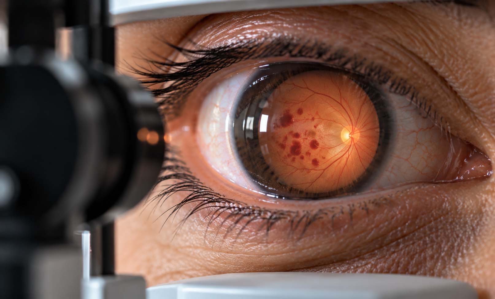
Diabetic retinopathy is a complication of diabetes that affects the retina, the light-sensitive tissue at the back of the eye responsible for detecting light and transmitting visual information to the brain.
Over time, elevated blood glucose levels can damage the retina's small blood vessels. These vessels may become weakened, leak fluid or blood, or become blocked, disrupting the retina's normal function.
As the disease progresses, abnormal new blood vessels may develop, increasing the risk of bleeding, scar tissue formation, and severe vision loss.
Diabetic retinopathy can affect people with either type 1 or type 2 diabetes. It may develop without noticeable symptoms, particularly during the early stages.
The condition is commonly classified into two main stages:
Non-Proliferative Diabetic Retinopathy (NPDR): The earlier stage, characterized by abnormalities in existing retinal blood vessels. Severity ranges from mild to severe.
Proliferative Diabetic Retinopathy (PDR): The more advanced stage, in which abnormal new blood vessels grow on the retina or optic disc. These fragile vessels can bleed and may lead to serious complications.
Diabetic Macular Edema (DME), which involves swelling of the central retina, can occur at different stages of diabetic retinopathy and may significantly affect central vision.
With regular screening, appropriate diabetes management, and timely ophthalmic treatment, many cases of severe vision loss can be prevented.
Symptoms
Diabetic retinopathy often causes no noticeable symptoms during its early stages.
As the condition progresses, possible symptoms include:
• Blurred or fluctuating vision.
• Difficulty reading or seeing fine details.
• Dark spots or floaters in the field of vision.
• Distorted central vision.
• Areas of missing or reduced vision.
• Reduced color clarity.
• Sudden loss of vision in advanced cases.
Symptoms may affect one or both eyes and can vary depending on the severity and location of retinal damage.
Sudden vision loss, a shower of new floaters, flashes of light, or a dark shadow across the vision requires urgent ophthalmic assessment.
Importantly, normal vision does not rule out diabetic retinopathy. Regular retinal examinations remain necessary even when no symptoms are present.
Causes
Diabetic retinopathy develops primarily as a result of long-term damage to the retinal microvascular system associated with diabetes.
Persistently elevated blood glucose levels can weaken and damage the small blood vessels supplying the retina.
These changes may result in:
• Leakage of fluid and blood into retinal tissue.
• Blockage of small retinal blood vessels.
• Reduced oxygen delivery to parts of the retina.
• Increased production of vascular endothelial growth factor (VEGF).
• Growth of abnormal, fragile blood vessels in advanced disease.
Fluid accumulation in the macula may cause diabetic macular edema, while fragile new blood vessels can lead to vitreous hemorrhage and scar tissue formation.
The severity and progression of diabetic retinopathy are influenced by metabolic control, the duration of diabetes, and other cardiovascular risk factors.
Risk Factors
Several factors increase the risk of developing or progressing diabetic retinopathy:
• Longer duration of diabetes.
• Persistently elevated blood glucose levels.
• High blood pressure.
• Abnormal blood lipid levels.
• Existing diabetic kidney disease.
• Pregnancy in women with pre-existing diabetes.
• Previous diabetic retinopathy.
• Inconsistent diabetes management or missed retinal follow-up appointments.
Both type 1 and type 2 diabetes can lead to diabetic retinopathy.
People with well-controlled diabetes may still develop retinal changes, which is why routine screening remains important.
Diagnosis
Diabetic retinopathy is diagnosed through a detailed examination of the retina, often supported by specialized retinal imaging.
1. Comprehensive Dilated Eye Examination
Dilating eye drops allow the ophthalmologist to examine the retina and identify bleeding, microaneurysms, swelling, abnormal blood vessels, and other retinal changes.
2. Fundus Photography
Digital retinal photographs help document diabetic retinal changes and monitor progression over time.
3. Optical Coherence Tomography (OCT)
OCT produces detailed cross-sectional images of the retina and is particularly useful for detecting and monitoring diabetic macular edema.
4. OCT Angiography (OCTA)
OCTA can provide additional information about retinal microvascular circulation without the use of an injected contrast dye.
5. Fluorescein Angiography (FFA)
In selected cases, fluorescein angiography helps identify leaking blood vessels, areas of reduced retinal blood flow, and abnormal vascular growth.
6. Visual Acuity Assessment
Vision testing measures how clearly the patient sees and helps evaluate the functional effect of retinal disease.
Not every patient requires every test. The ophthalmologist selects investigations based on the clinical findings and stage of diabetic retinopathy.
Treatment Options
Treatment depends on the severity of diabetic retinopathy, the presence of diabetic macular edema, and the risk of progressive vision loss.
The main objectives are to preserve vision, reduce retinal complications, and prevent further damage.
1. Diabetes and Cardiovascular Risk Management
Effective management of blood glucose, blood pressure, and blood lipid levels is an essential part of diabetic retinopathy care.
Patients should work closely with their diabetes care team to maintain individualized treatment targets.
2. Observation and Regular Monitoring
Mild non-proliferative diabetic retinopathy may not require immediate ophthalmic treatment.
Instead, the ophthalmologist may recommend regular retinal examinations and imaging to monitor the condition.
Follow-up frequency depends on the severity of retinal findings and the individual's risk of progression.
3. Intravitreal Anti-VEGF Injections
Anti-VEGF medications are injected into the eye to reduce abnormal blood vessel activity and retinal fluid leakage.
They are commonly used to treat diabetic macular edema that affects vision and may also be used in selected patients with proliferative diabetic retinopathy.
Multiple injections and ongoing monitoring may be necessary.
4. Retinal Laser Treatment
Laser photocoagulation can help manage particular forms of diabetic retinal disease.
Panretinal photocoagulation (PRP) is an established treatment for proliferative diabetic retinopathy. It reduces the stimulus for abnormal blood vessel growth and helps lower the risk of severe vision loss.
Focal or grid laser treatment may be appropriate for selected patients with diabetic macular edema.
5. Intravitreal Corticosteroid Treatment
Steroid injections or implants may be considered for selected cases of diabetic macular edema, particularly when other treatment options are unsuitable or response is inadequate.
Potential complications include increased eye pressure and cataract progression.
6. Vitrectomy Surgery
Vitrectomy may be required in advanced cases, including certain patients with non-clearing vitreous hemorrhage or tractional retinal detachment.
During surgery, the vitreous gel and associated blood or scar tissue may be removed to manage retinal complications.
Treatment decisions are individualized by a retinal specialist. Some treatments can improve vision, but recovery depends on the extent of existing retinal damage.
Prevention & Care
Diabetic retinopathy cannot always be prevented, but the risk of progression and severe vision loss can often be reduced.
Recommended measures include:
• Maintain blood glucose levels as close as safely possible to individualized targets.
• Monitor and manage blood pressure and blood lipid levels.
• Attend regular diabetic retinal screening and dilated eye examinations.
• Follow the treatment plan recommended by the diabetes care team.
• Avoid smoking and maintain a healthy lifestyle.
• Attend all scheduled retinal follow-up appointments, even when vision seems normal.
• Report new visual symptoms promptly.
• Inform the ophthalmologist when planning pregnancy or becoming pregnant if you have pre-existing diabetes.
When should eye examinations begin?
For adults with type 1 diabetes, an initial comprehensive dilated eye examination is generally recommended within five years after diagnosis.
For people with type 2 diabetes, the initial examination should be performed at the time of diagnosis.
Subsequent examinations are commonly performed annually, although the interval may be adjusted according to retinal findings, diabetes control, and individual risk.
Patients with progressing or sight-threatening disease may need substantially more frequent examinations.
Pregnant patients with pre-existing diabetes require individualized retinal assessment and monitoring.
When should you seek urgent medical attention?
Seek urgent ophthalmic assessment for sudden loss of vision, significant new floaters, flashes of light, or a shadow across the visual field.
Early assessment can help identify complications requiring prompt treatment.
Frequently Asked Questions
What is diabetic retinopathy?
Can diabetic retinopathy develop without symptoms?
Can people with type 2 diabetes develop diabetic retinopathy?
Can diabetic retinopathy cause blindness?
Is diabetic retinopathy reversible?
What is diabetic macular edema?
How often should someone with diabetes have an eye examination?
Are eye injections necessary for every patient?
Can retinal laser treatment restore normal vision?
Can diabetic retinopathy be prevented?
Is diabetic retinopathy treatment lifelong?
Our team can help you with an individual medical assessment.