
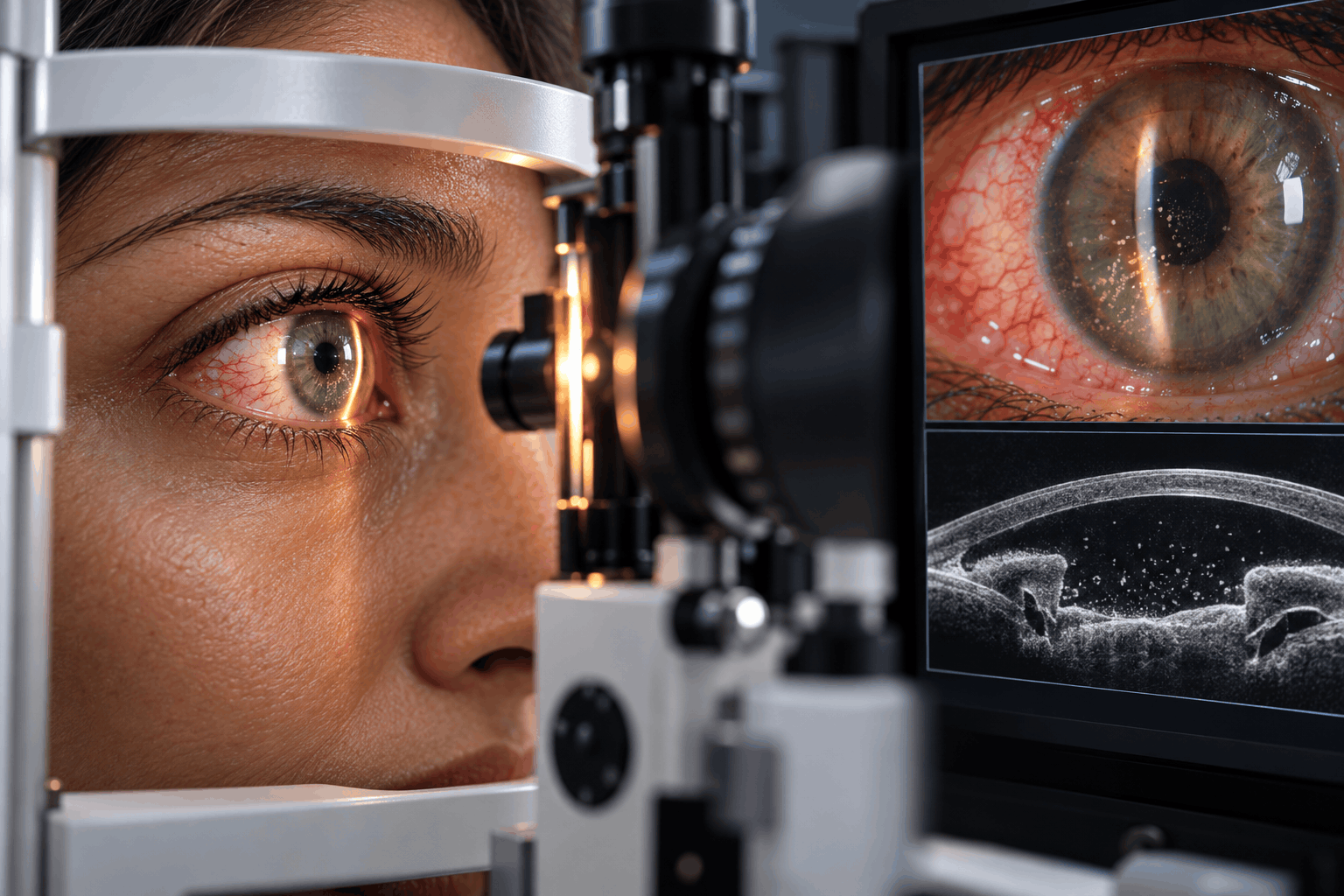
What Is It?
Uveitis refers to a group of inflammatory conditions affecting the inside of the eye, particularly the uvea, the middle layer of the eye. The uvea consists of three main structures: the iris, the ciliary body, and the choroid.
The iris controls the amount of light entering the eye, the ciliary body contributes to focusing and fluid production, and the choroid supplies oxygen and nutrients to the outer retina.
Inflammation may affect one or more of these structures and can also involve nearby tissues, including the vitreous, retina, and optic nerve.
Uveitis may develop suddenly or gradually. Some patients experience a single episode, while others develop recurrent or persistent inflammation.
Types of Uveitis
Anterior Uveitis
The most common form, anterior uveitis primarily affects the iris and sometimes the ciliary body. It is also known as iritis or iridocyclitis. Symptoms often include eye pain, redness, blurred vision, and sensitivity to light.
Intermediate Uveitis
This form mainly involves the vitreous and peripheral retina. Patients may notice floaters and blurred vision, sometimes without significant redness or pain.
Posterior Uveitis
Posterior uveitis involves the choroid, retina, or both. It may affect central vision and can cause floaters, blurred vision, or visual disturbances. The eye may appear externally normal despite significant inflammation.
Panuveitis
Panuveitis involves inflammation affecting the anterior, intermediate, and posterior segments of the eye. It may be associated with systemic inflammatory diseases, infections, or other conditions.
Uveitis is also classified by its duration and course, including acute, recurrent, and chronic forms.
Although some episodes respond well to treatment, untreated or recurrent uveitis can lead to complications such as cataracts, glaucoma, macular edema, retinal damage, and permanent vision loss.
Early ophthalmic assessment is essential because the management of uveitis depends on the underlying cause, anatomical location, and severity of inflammation.
Symptoms
Symptoms of uveitis vary depending on which part of the eye is affected and how quickly inflammation develops.
Common symptoms include:
• Eye redness, particularly around the iris.
• Eye pain or deep aching discomfort.
• Sensitivity to light (photophobia).
• Blurred or reduced vision.
• Floaters, appearing as small moving dots, shadows, or threads in the visual field.
• Excessive tearing or watery eyes.
• Difficulty focusing clearly on objects.
• Visual haze or reduced contrast.
• Changes in the appearance or shape of the pupil in certain cases.
Anterior Uveitis Symptoms
Anterior uveitis commonly produces redness, pain, photophobia, and blurred vision. Symptoms may appear suddenly over hours or days.
Intermediate Uveitis Symptoms
Patients often experience floaters and blurred vision. Eye pain and visible redness may be absent.
Posterior Uveitis Symptoms
Blurred or distorted vision, floaters, and areas of reduced vision may occur. Some patients have little external redness or discomfort.
Panuveitis Symptoms
Symptoms may include a combination of pain, redness, floaters, photophobia, and significant visual impairment.
In some cases, particularly in children or patients with chronic inflammation, uveitis may develop with few noticeable symptoms.
Symptoms can affect one eye or both eyes, either simultaneously or at different times.
Any new eye pain, marked light sensitivity, unexplained blurred vision, or sudden onset of floaters should prompt urgent ophthalmic assessment.
Sudden severe vision loss requires immediate emergency care.
Because symptoms can resemble other eye conditions, a complete examination is necessary to confirm the diagnosis.
Causes
Uveitis develops when inflammatory processes affect tissues inside the eye. The inflammation may be associated with immune-system disorders, infections, injury, or other ocular conditions.
In many patients, no definite underlying cause is identified. Such cases are described as idiopathic uveitis.
1. Noninfectious Immune-Mediated Conditions
The immune system may mistakenly trigger inflammation within ocular tissues.
Uveitis can occur in association with systemic inflammatory diseases, including:
• Ankylosing spondylitis and other HLA-B27-associated spondyloarthropathies.
• Behçet disease.
• Sarcoidosis.
• Juvenile idiopathic arthritis.
• Inflammatory bowel disease.
• Psoriatic arthritis.
• Vogt–Koyanagi–Harada disease.
• Certain other autoimmune or inflammatory disorders.
These conditions may affect the joints, skin, blood vessels, lungs, gastrointestinal system, or other organs in addition to the eyes.
2. Infectious Causes
Some forms of uveitis are caused by infectious microorganisms or immune responses associated with infection.
Examples include:
• Herpes simplex virus.
• Varicella-zoster virus.
• Cytomegalovirus in selected patients.
• Toxoplasma gondii.
• Syphilis.
• Tuberculosis.
• Other bacterial, viral, fungal, or parasitic infections, depending on exposure and regional prevalence.
Correct identification of an infection is essential because treatment differs from that used for noninfectious inflammation.
3. Eye Trauma
Blunt or penetrating eye injuries may trigger inflammation involving internal ocular structures.
4. Postoperative Inflammation
Inflammation may develop after certain eye procedures. The ophthalmologist must distinguish expected postoperative inflammation from infection or other complications.
5. Medication-Associated Uveitis
Certain medications may rarely cause intraocular inflammation. A detailed medication history can help identify possible associations.
6. Masquerade Conditions
Occasionally, disorders such as intraocular lymphoma may resemble uveitis. These conditions require specific diagnostic investigation.
Uveitis is not always contagious. Most noninfectious forms cannot spread from one person to another.
The cause determines the treatment strategy, so an ophthalmologist may recommend additional medical investigations when appropriate.
Risk Factors
Uveitis can affect people of different ages and backgrounds, and it may occur without any recognized risk factors.
However, several conditions and circumstances are associated with an increased likelihood of developing certain forms of uveitis.
Age
Uveitis commonly affects adults, including those between approximately 20 and 60 years of age, although children and older adults can also develop the condition.
Autoimmune and Inflammatory Diseases
People with conditions such as ankylosing spondylitis, Behçet disease, sarcoidosis, or juvenile idiopathic arthritis may be at increased risk.
HLA-B27 Association
The HLA-B27 genetic marker is associated with certain forms of acute anterior uveitis, particularly in patients with spondyloarthritis. Having this marker does not necessarily mean uveitis will develop.
Previous Uveitis
A history of uveitis increases the possibility of future episodes, particularly in recurrent inflammatory conditions.
Certain Infections
Previous or active infections, including herpes viruses, toxoplasmosis, tuberculosis, and syphilis, may be associated with specific forms of intraocular inflammation.
Eye Trauma or Surgery
Injury to the eye or certain postoperative complications may trigger inflammatory responses.
Immunosuppression
Some infections affecting the eye are more likely in patients with weakened immune systems.
Smoking
Tobacco smoking has been associated with an increased risk of uveitis and may contribute to poorer ocular inflammatory outcomes.
Systemic Symptoms
Joint pain, skin changes, oral ulcers, gastrointestinal symptoms, or respiratory symptoms may indicate an associated systemic disorder, depending on the overall clinical picture.
The presence of a risk factor does not confirm the diagnosis. Clinical examination and targeted investigations are necessary to determine whether uveitis is present and identify its cause.
Diagnosis
Uveitis is diagnosed through a detailed ophthalmic examination. Additional laboratory or imaging investigations may be required to determine the cause, assess the extent of inflammation, and identify associated complications.
1. Medical and Ocular History
The ophthalmologist asks about the onset, duration, and recurrence of symptoms, previous eye disease, trauma, surgery, infections, medications, and systemic inflammatory conditions.
Associated symptoms such as joint pain, mouth ulcers, skin rashes, back pain, respiratory complaints, and gastrointestinal problems may provide diagnostic clues.
2. Visual Acuity Testing
Visual acuity is measured to assess the effect of inflammation on central vision and provide a baseline for monitoring treatment.
3. Slit-Lamp Examination
A slit-lamp microscope allows detailed examination of the front structures of the eye.
The ophthalmologist looks for inflammatory cells and flare in the anterior chamber, corneal deposits, changes in the iris, and other signs of inflammation.
4. Intraocular Pressure Measurement
Eye pressure is measured because uveitis and certain treatments can cause pressure changes, increasing the risk of glaucoma or other complications.
5. Dilated Fundus Examination
Pupil dilation enables examination of the vitreous, retina, macula, choroid, and optic nerve.
This is particularly important when intermediate, posterior, or panuveitis is suspected.
6. Optical Coherence Tomography (OCT)
OCT provides detailed cross-sectional images of the retina and is useful for detecting complications such as macular edema and monitoring structural changes.
7. Fundus Photography
Retinal photographs may be used to document inflammatory lesions and assess changes over time.
8. Fluorescein or Indocyanine Green Angiography
In selected cases, specialized imaging may help identify retinal vascular leakage, inflammatory changes, or choroidal involvement.
9. Laboratory Investigations
Blood tests and infectious disease screening may be requested according to the type of uveitis, patient history, and clinical findings.
Tests may evaluate possible infections, systemic inflammatory conditions, or other associated disorders.
Broad laboratory testing is not required for every patient; investigations should be tailored to the clinical presentation.
10. Additional Medical Assessment
Some patients may be referred to a rheumatologist, infectious disease specialist, or other physician when a systemic condition is suspected.
11. Ocular Fluid Analysis
In selected cases, samples of aqueous or vitreous fluid may be obtained for specialized tests, including polymerase chain reaction (PCR), to investigate infections or other causes.
The diagnostic approach is individualized. Regular follow-up is important because the appearance and severity of uveitis can change during treatment.
Treatment Options
Treatment of uveitis aims to control intraocular inflammation, relieve symptoms, address any underlying cause, prevent recurrence where possible, and preserve vision.
The treatment plan depends on the location and severity of inflammation, whether infection is present, and the patient's overall medical condition.
1. Corticosteroid Eye Drops
Prescription corticosteroid drops are commonly used to treat noninfectious anterior uveitis.
These medications reduce inflammation and help relieve symptoms.
The dosage and duration are determined by the ophthalmologist and adjusted according to the response to treatment.
Steroid drops must not be used without medical supervision because they can worsen certain infections and increase eye pressure.
2. Cycloplegic and Pupil-Dilating Drops
These drops may be prescribed for anterior uveitis to relax the muscles inside the eye, relieve pain caused by muscle spasm, and reduce the risk of certain iris-related complications.
Temporary blurred near vision and light sensitivity may occur during use.
3. Periocular or Intraocular Corticosteroid Therapy
Some patients with intermediate or posterior noninfectious uveitis may require corticosteroid injections around or inside the eye.
Sustained-release steroid implants may be considered in selected cases requiring longer-term local treatment.
These therapies carry risks, including increased intraocular pressure and cataract development.
4. Systemic Corticosteroids
Oral or intravenous corticosteroids may be required for severe noninfectious uveitis, especially when inflammation affects the back of the eye or both eyes.
Systemic treatment requires careful supervision because corticosteroids can cause significant adverse effects.
5. Immunomodulatory Therapy
For recurrent, chronic, severe, or steroid-dependent noninfectious uveitis, medications that regulate immune activity may be necessary.
Examples include methotrexate, mycophenolate mofetil, azathioprine, and other specialist-prescribed therapies.
Treatment may be coordinated with a rheumatologist or another relevant specialist.
6. Biological Therapies
In selected cases of noninfectious uveitis, biological medications such as adalimumab may be considered when conventional treatments are insufficient or unsuitable.
These medications require assessment for potential infections and appropriate ongoing monitoring.
7. Treatment of Infectious Uveitis
When uveitis is caused by an infection, treatment must target the responsible microorganism.
Depending on the diagnosis, therapy may include antiviral, antibiotic, antifungal, or antiparasitic medication.
Anti-inflammatory treatment may sometimes be used alongside antimicrobial therapy under specialist supervision.
Immunosuppressive treatment should not be started without adequately evaluating possible infectious causes.
8. Management of Eye Pressure
Uveitis or corticosteroid therapy can lead to elevated eye pressure.
Additional medications may be needed to protect the optic nerve and reduce the risk of glaucoma.
9. Treatment of Associated Complications
Macular edema, cataracts, glaucoma, vitreous opacities, or retinal complications may require specific medical, laser, or surgical treatment.
10. Surgical Management
Surgery is not routinely required for uncomplicated uveitis.
However, procedures such as cataract surgery, glaucoma surgery, or pars plana vitrectomy may be appropriate in selected patients to manage complications or assist diagnosis.
Whenever possible, inflammation is controlled before elective intraocular surgery.
11. Monitoring and Long-Term Follow-Up
Regular examinations are essential to assess inflammatory activity, visual acuity, intraocular pressure, treatment response, and medication side effects.
Some patients need treatment for only a limited period, while others require long-term management.
Patients should not stop or change prescribed medication without consulting their ophthalmologist because premature discontinuation may allow inflammation to return.
With timely diagnosis and individualized management, many patients achieve good control of inflammation and preserve useful vision.
Prevention & Care
Not all cases of uveitis can be prevented because the underlying cause is often unknown.
However, several measures can reduce the risk of complications, support long-term eye health, and help identify recurrences early.
• Attend regular ophthalmic examinations when diagnosed with uveitis.
• Seek prompt medical assessment for new eye pain, redness, sensitivity to light, floaters, or blurred vision.
• Follow prescribed treatment schedules carefully.
• Do not stop corticosteroid or other anti-inflammatory medication without medical advice.
• Monitor eye pressure when receiving corticosteroid treatment.
• Follow the recommended schedule for retinal examination and OCT imaging when indicated.
• Inform the ophthalmologist about any history of autoimmune disease, infection, or recurrent eye inflammation.
• Maintain appropriate treatment for associated systemic inflammatory conditions.
• Attend specialist follow-up appointments when receiving immunosuppressive or biological therapies.
• Avoid smoking and seek support for smoking cessation when appropriate.
• Wear protective eyewear during activities that may cause ocular injury.
• Report any significant medication side effects promptly.
• Keep a record of previous uveitis episodes and treatment when possible.
Patients with recurrent uveitis should learn to recognize their individual warning symptoms.
However, absence of pain or redness does not always exclude active inflammation, particularly in intermediate or posterior uveitis.
Children with certain autoimmune conditions may need regular screening even if they do not complain of eye symptoms.
Prompt treatment of infectious causes and appropriate management of immune-related disorders may reduce the risk of lasting ocular damage.
Seek urgent ophthalmic assessment for new or worsening vision changes, severe light sensitivity, unexplained eye pain, or a markedly red eye.
Sudden significant vision loss requires emergency care.
Because untreated inflammation can affect the retina, optic nerve, and other structures, long-term follow-up remains important even after symptoms improve.
Frequently Asked Questions
What is uveitis?
What are the main symptoms of uveitis?
Is uveitis a serious eye condition?
What are the different types of uveitis?
What causes uveitis?
Is uveitis contagious?
Can uveitis affect both eyes?
Can uveitis develop without eye pain?
How is uveitis diagnosed?
Does every patient with uveitis need blood tests?
Can uveitis be related to autoimmune disease?
How is uveitis treated?
Are steroid eye drops safe for uveitis?
Can uveitis return after treatment?
Can uveitis cause glaucoma or cataracts?
Is surgery always necessary for uveitis?
Can uveitis cause permanent vision loss?
Can children develop uveitis?
How long does uveitis take to heal?
When should I seek urgent eye care?
Our team can help you with an individual medical assessment.