
What Is It?
Retinal artery occlusion (RAO) is a serious vascular condition in which blood flow through an artery supplying the retina becomes blocked.
The retina is the light-sensitive tissue at the back of the eye. It requires a continuous supply of oxygen and nutrients to maintain normal vision.
When retinal arterial blood flow is interrupted, retinal cells become deprived of oxygen. Prolonged interruption can cause retinal infarction and permanent visual damage.
There are two major forms:
1. Central Retinal Artery Occlusion (CRAO)
CRAO occurs when the main artery supplying much of the inner retina becomes obstructed.
It typically causes sudden, severe, painless loss of vision in one eye.
Acute CRAO with retinal infarction is considered a form of ischemic stroke and requires emergency medical assessment.
2. Branch Retinal Artery Occlusion (BRAO)
BRAO occurs when one of the smaller branches of the retinal arterial circulation becomes blocked.
Vision loss may affect only a particular area of the visual field, depending on the location of the occlusion.
Although the visual impact may be less extensive than with CRAO, acute symptomatic BRAO also requires urgent assessment for potential stroke-related causes.
A less common condition, ophthalmic artery occlusion, affects a larger arterial supply to the eye and can produce profound visual impairment.
Retinal artery occlusion is often associated with an embolus, such as material originating from a carotid artery plaque or the heart.
Occasionally, inflammation of blood vessels, particularly giant cell arteritis in older adults, is responsible.
Retinal artery occlusion is not the same as retinal vein occlusion. Arterial occlusion interrupts the delivery of oxygenated blood, whereas venous occlusion affects drainage of blood from the retina.
Because RAO may indicate an increased risk of brain stroke or other cardiovascular events, urgent systemic assessment is essential even when visual symptoms are isolated.
Established retinal infarction can cause irreversible visual loss. Rapid recognition, appropriate emergency evaluation, and management of underlying vascular risks are therefore critical.
Symptoms
Retinal artery occlusion usually produces sudden visual symptoms, most often affecting one eye.
Common symptoms include:
• Sudden, painless loss of vision in one eye.
• Severe blurring or darkening of vision.
• Sudden loss of part of the visual field.
• A fixed dark or missing area of vision.
• Difficulty recognizing objects or reading because of sudden visual impairment.
In CRAO, loss of vision is often profound because a large area of the retina is affected.
In BRAO, patients may notice a localized visual field defect rather than complete loss of central vision.
Symptoms usually develop within seconds or minutes.
Some patients experience temporary episodes of visual loss before a permanent event. This can represent transient retinal ischemia and requires emergency stroke evaluation.
IMPORTANT — MEDICAL EMERGENCY
Sudden painless loss of vision, whether partial or complete, requires immediate assessment at an emergency department equipped to evaluate stroke.
Do not wait for the vision to return.
Do not delay care to arrange a routine ophthalmology appointment.
Even temporary vision loss in one eye can be a warning sign of a serious vascular problem.
Other stroke symptoms may include facial weakness, arm weakness, speech difficulty, or problems with balance, but retinal artery occlusion can occur without these symptoms.
In adults over 50, new headache, scalp tenderness, jaw pain while chewing, fever, or shoulder and hip stiffness accompanying visual symptoms may raise concern for giant cell arteritis, another emergency requiring immediate medical attention.
Causes
Retinal artery occlusion develops when blood flow through a retinal artery becomes critically reduced or blocked.
Possible mechanisms include:
1. Embolism
An embolus is material that travels through the bloodstream and becomes lodged in a smaller blood vessel.
Emboli may originate from atherosclerotic plaques in the carotid arteries or from the heart.
Cardiac conditions such as atrial fibrillation or certain heart valve disorders can contribute to embolic risk.
2. Atherosclerotic Vascular Disease
Atherosclerosis involves the accumulation of plaque within blood vessel walls.
It can contribute to embolism or impaired blood circulation.
3. Thrombosis
In selected patients, a blood clot forms within an artery and obstructs circulation.
4. Giant Cell Arteritis
Giant cell arteritis is an inflammatory disease affecting medium and large arteries, usually in adults over 50.
It can interrupt blood supply to the eye and cause sudden vision loss.
Suspected giant cell arteritis requires immediate medical assessment and urgent treatment because the other eye and overall health may be at risk.
5. Other Vascular or Inflammatory Disorders
Less common causes include certain vasculitic conditions, vascular abnormalities, and disorders affecting blood clotting.
These may be considered particularly in younger patients or when the presentation is unusual.
6. Rare Procedure-Related or Traumatic Events
Retinal arterial occlusion may occasionally occur in association with specific medical procedures, trauma, or other uncommon circumstances.
Regardless of the cause, prolonged reduction in blood supply may lead to retinal ischemia and irreversible damage.
Identifying the source of the obstruction is essential not only for eye care but also for preventing future vascular events.
Risk Factors
Several factors are associated with retinal artery occlusion, particularly when the cause is embolic or atherosclerotic.
Important risk factors include:
• Increasing age.
• High blood pressure.
• Diabetes mellitus.
• Elevated cholesterol or other abnormal blood lipid levels.
• Smoking.
• Carotid artery atherosclerosis.
• Atrial fibrillation and certain heart diseases.
• Previous stroke or transient ischemic attack.
• Established cardiovascular or peripheral vascular disease.
Other important considerations include:
• Giant cell arteritis, particularly in adults over 50.
• Certain inflammatory or autoimmune vascular disorders.
• Selected blood clotting disorders.
• Uncommon vascular abnormalities.
Retinal artery occlusion may also occur in younger individuals, sometimes requiring investigation for less common causes.
The presence of a risk factor does not mean that retinal artery occlusion will necessarily develop.
An episode of RAO may be the first indication of an underlying cardiovascular condition.
Every patient requires an individualized assessment to identify possible embolic, inflammatory, or vascular causes and reduce the risk of future events.
Diagnosis
Retinal artery occlusion is diagnosed through urgent clinical assessment, a detailed eye examination, and investigations for associated stroke and cardiovascular disease.
Emergency evaluation must not be delayed while waiting for outpatient retinal imaging.
1. Immediate Emergency Assessment
Patients with new, sudden vision loss require urgent assessment in a stroke-capable emergency department.
The medical team determines when symptoms began and evaluates eligibility for time-sensitive emergency interventions.
2. Medical and Vascular History
The doctor reviews the onset of visual symptoms, previous transient visual loss, hypertension, diabetes, cardiovascular disease, smoking history, and medication use.
In older patients, symptoms suggesting giant cell arteritis are specifically assessed.
3. Visual Acuity and Pupillary Examination
Visual acuity testing measures the extent of visual loss.
Pupillary responses may reveal a relative afferent pupillary defect when retinal damage is substantial.
4. Dilated Fundus Examination
The ophthalmologist examines the retinal arteries, macula, and optic nerve.
Findings may include retinal whitening, attenuated arteries, visible embolic material, or a characteristic cherry-red spot in some patients with CRAO.
Very early retinal artery occlusion may not show all of these changes.
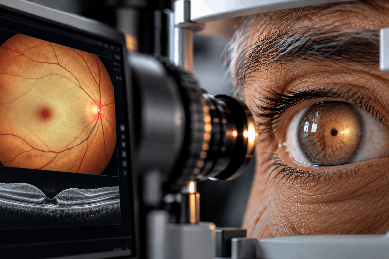
5. Optical Coherence Tomography (OCT)
OCT can demonstrate changes in the inner retinal layers caused by acute ischemia.
It is also useful in evaluating retinal damage over time.
6. Fundus Photography and Retinal Vascular Imaging
Fundus photographs document the retinal appearance.
Fluorescein angiography or OCT angiography may be used selectively to assess retinal circulation.
These tests should not delay urgent systemic evaluation.
7. Stroke and Cardiovascular Investigations
Emergency investigations are selected by the stroke team and may include:
• Brain imaging.
• Carotid and other vascular imaging.
• Electrocardiography.
• Cardiac monitoring.
• Echocardiography when indicated.
• Blood glucose and lipid assessment.
• Additional laboratory investigations based on the clinical situation.
8. Evaluation for Giant Cell Arteritis
In patients over 50 with concerning clinical findings, investigations may include erythrocyte sedimentation rate (ESR), C-reactive protein (CRP), platelet count, and other specialist tests.
When giant cell arteritis is strongly suspected, urgent treatment should not be delayed while awaiting confirmatory testing.
Not every patient requires every investigation.
Diagnosis and management require coordination between emergency medicine, stroke specialists, ophthalmologists, and other relevant medical teams.
Treatment Options
Retinal artery occlusion is a medical emergency, particularly when visual symptoms have started suddenly.
Management has three major goals:
• Identify any possibility of time-sensitive treatment.
• Investigate the cause and reduce the risk of brain stroke or other vascular events.
• Monitor and manage ocular complications.
1. Immediate Referral to a Stroke-Capable Emergency Department
Patients with acute symptomatic CRAO or BRAO should receive emergency stroke assessment.
Time is critical because the opportunity for certain interventions, if appropriate, may be limited.
Patients should not wait for a routine ophthalmology clinic appointment.
2. Evaluation for Acute Stroke Treatment
In selected patients with acute CRAO, intravenous thrombolysis may be considered by an experienced stroke team when the patient presents within an appropriate time window and meets established eligibility and safety criteria.
However, the evidence for visual benefit remains uncertain, and thrombolysis is not routine treatment for every patient with retinal artery occlusion.
The decision must be made by the treating emergency and stroke specialists after consideration of potential benefits, including the risk of serious bleeding.
Other proposed acute treatments have not been established as reliably effective in restoring vision.
3. Treatment of Giant Cell Arteritis
When giant cell arteritis is strongly suspected or diagnosed, urgent systemic corticosteroid treatment is generally required under specialist supervision.
The primary aims include protecting the other eye and reducing serious systemic complications.
Treatment should not be delayed for confirmatory test results when clinical suspicion is high.
4. Cardiovascular and Stroke Risk Management
After emergency assessment, treatment focuses on the identified vascular risk factors and embolic sources.
Depending on the cause, management may include:
• Treatment of high blood pressure.
• Diabetes management.
• Cholesterol-lowering therapy.
• Smoking cessation.
• Appropriate antiplatelet or anticoagulant therapy for specific medical indications.
• Management of atrial fibrillation or other cardiac disease.
• Evaluation and management of significant carotid artery disease.
Medication choices are determined by the medical and stroke teams, not by the retinal findings alone.
5. Retinal Monitoring and Treatment of Complications
Patients may require ophthalmic follow-up to monitor for:
• Persistent visual impairment.
• Retinal ischemic changes.
• Abnormal new blood vessel growth.
• Neovascular glaucoma.
In patients who develop retinal or iris neovascularization, retinal laser treatment such as panretinal photocoagulation may be recommended.
Additional therapies may be necessary depending on the complication.
Laser treatment is not a method of reopening the original blocked retinal artery.
6. Low Vision Rehabilitation
If permanent visual impairment remains, support may include:
• Low vision assessment.
• Magnification devices.
• Electronic reading aids.
• Adaptive lighting.
• Practical rehabilitation for daily activities.
7. Treatments Without Established Routine Benefit
Traditional interventions such as ocular massage, anterior chamber paracentesis, or attempts to rapidly lower eye pressure have not demonstrated reliable visual benefit in acute CRAO and may carry risks.
They should not delay emergency stroke care.
Hyperbaric oxygen therapy has been investigated, but its role remains uncertain and availability is limited. It is not established as standard treatment for every RAO patient.
Visual recovery varies according to the type, severity, and duration of retinal ischemia.
Once retinal infarction has occurred, vision loss is often permanent. Rapid evaluation is essential both for potential acute treatment and for prevention of future stroke or cardiovascular events.
Prevention & Care
Not every retinal artery occlusion can be prevented, but management of cardiovascular risk factors may reduce the likelihood of future vascular events.
Recommended measures include:
• Monitor and control blood pressure.
• Manage diabetes according to individualized medical targets.
• Treat elevated cholesterol and other cardiovascular risk factors.
• Avoid smoking.
• Maintain regular physical activity as medically appropriate.
• Follow prescribed treatment for heart rhythm abnormalities or vascular disease.
• Attend recommended cardiovascular and ophthalmic follow-up appointments.
• Take prescribed antiplatelet or anticoagulant medications only when directed by the treating physician.
• Report transient episodes of vision loss even when vision returns.
Why Is General Medical Evaluation Important?
Retinal artery occlusion can indicate disease affecting blood vessels elsewhere in the body.
Patients may be at increased risk of brain stroke or other cardiovascular events.
The stroke and medical teams assess whether additional treatment is needed to reduce future risk.
When Should You Seek Emergency Care?
Seek emergency medical assistance immediately for:
• Sudden loss of vision in one eye.
• Sudden loss of part of the visual field.
• Temporary complete or partial vision loss.
• Sudden visual symptoms accompanied by facial weakness, arm weakness, or speech difficulty.
Do not drive yourself if you experience sudden visual loss or other possible stroke symptoms.
Do not wait for symptoms to resolve or for an available routine eye appointment.
In patients over 50, sudden visual symptoms associated with new headache, scalp tenderness, or jaw pain while chewing require urgent evaluation for giant cell arteritis.
Follow-Up After Retinal Artery Occlusion
Follow-up may involve an ophthalmologist, neurologist, cardiologist, or primary care physician.
The medical team may recommend ongoing monitoring of cardiovascular risk factors and eye examinations for complications.
Patients with permanent visual impairment may also benefit from low vision rehabilitation.
Early recognition, emergency assessment, and prevention of further vascular events are the most important aspects of care.
Frequently Asked Questions
What is retinal artery occlusion?
Is retinal artery occlusion a medical emergency?
What is the difference between CRAO and BRAO?
Is central retinal artery occlusion considered a stroke?
Does retinal artery occlusion cause pain?
What are the first symptoms of retinal artery occlusion?
Can retinal artery occlusion affect both eyes?
What causes retinal artery occlusion?
Can giant cell arteritis cause retinal artery occlusion?
Can vision return after retinal artery occlusion?
Can eye injections cure retinal artery occlusion?
Is laser treatment used for retinal artery occlusion?
Can clot-dissolving medication treat CRAO?
Why do I need a stroke evaluation if the problem is in my eye?
Can retinal artery occlusion happen again?
Can high blood pressure increase the risk?
What is the difference between retinal artery and retinal vein occlusion?
Do I need blood-thinning medication?
What if my vision returns after a few minutes?
When should I go to the emergency department?
Our team can help you with an individual medical assessment.