
What Is It?
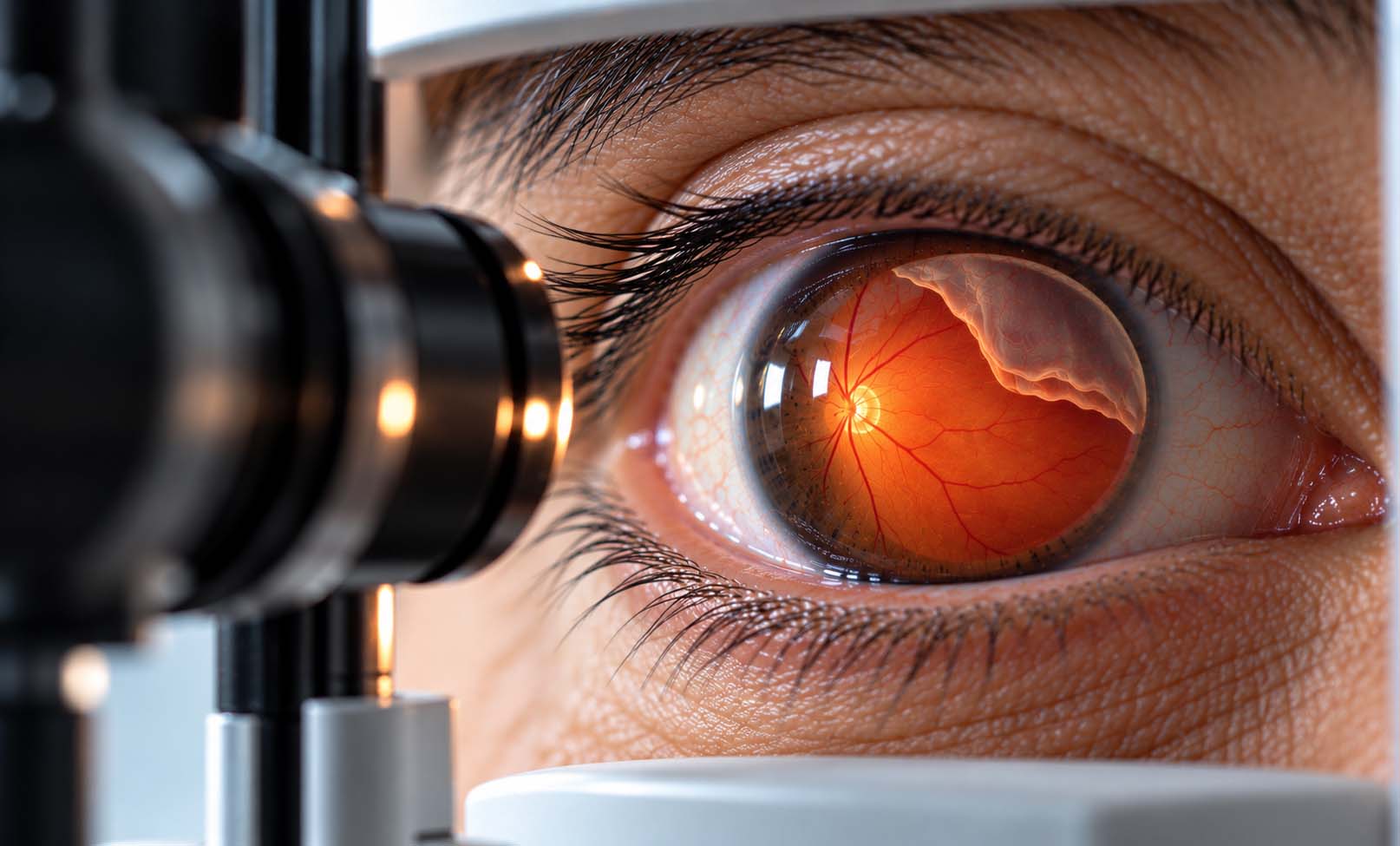
Retinal detachment occurs when the retina, the light-sensitive layer of tissue lining the back of the eye, separates from the tissue beneath it.
The retina converts incoming light into electrical signals that are transmitted to the brain through the optic nerve, allowing us to see.
When the retina becomes detached, its normal function is disrupted. Without prompt treatment, retinal cells may become permanently damaged, resulting in partial or severe vision loss.
Retinal detachment may affect a small area or extend across a larger portion of the retina. Vision loss can become more severe if the macula, the central part of the retina responsible for detailed vision, becomes detached.
There are three main types of retinal detachment:
1. Rhegmatogenous Retinal Detachment
The most common type. It occurs when a tear or hole develops in the retina, allowing fluid from inside the eye to pass underneath and separate the retina from the underlying tissue.
2. Tractional Retinal Detachment
This occurs when scar tissue pulls the retina away from its normal position. It is commonly associated with advanced proliferative diabetic retinopathy.
3. Exudative Retinal Detachment
This occurs when fluid accumulates beneath the retina without a retinal tear. Possible causes include inflammatory disorders, vascular abnormalities, and certain ocular tumors.
Retinal detachment is an ophthalmic emergency. Prompt specialist assessment and appropriate treatment are essential to protect remaining vision.
Symptoms
Retinal detachment is usually painless, and its symptoms often appear suddenly.
Warning signs include:
• A sudden increase in floaters, which may look like dark spots, threads, or cobwebs moving across the field of vision.
• Flashes of light, particularly in peripheral vision.
• A dark shadow or curtain moving across part of the visual field.
• Sudden blurred or reduced vision.
• Loss of peripheral (side) vision.
• Loss of central vision if the macula becomes involved.
Symptoms may initially affect only one eye.
A small retinal detachment may sometimes produce few or no noticeable symptoms.
IMPORTANT: SEEK IMMEDIATE EYE CARE
New flashes, a sudden shower of floaters, a dark curtain over vision, or sudden visual loss may indicate a retinal tear or detachment.
These symptoms require urgent examination by an ophthalmologist or evaluation at an emergency department, even if there is no pain.
Not every floater or flash means retinal detachment, but a dilated eye examination is necessary to distinguish harmless vitreous changes from a potentially sight-threatening retinal problem.
Causes
Retinal detachment occurs when the retina separates from its normal supporting tissue. The mechanism depends on the type of detachment.
1. Retinal Tears and Vitreous Changes
As people age, the vitreous gel inside the eye gradually changes and may separate from the retina. This process is called posterior vitreous detachment.
In most cases, posterior vitreous detachment does not cause retinal detachment. However, if the vitreous pulls strongly on the retina, it may produce a retinal tear.
Fluid can then pass through the tear and accumulate beneath the retina, resulting in rhegmatogenous retinal detachment.
2. Retinal Scar Tissue
Scar tissue forming on the retinal surface can contract and pull the retina away from the back of the eye.
This mechanism is known as tractional retinal detachment and is particularly associated with advanced diabetic retinal disease.
3. Fluid Accumulation Beneath the Retina
Certain inflammatory, vascular, or other ocular disorders may cause fluid to accumulate beneath the retina without a tear.
This can lead to exudative retinal detachment.
4. Eye Injury or Previous Surgery
Trauma and certain previous eye surgeries may increase the likelihood of retinal tears or detachment in susceptible individuals.
The cause must be identified because different types of retinal detachment require different treatment approaches.
Risk Factors
Several conditions and characteristics can increase the risk of retinal detachment:
• Increasing age and age-related vitreous changes.
• High myopia (severe nearsightedness).
• A previous retinal tear or retinal detachment.
• Retinal detachment in the other eye.
• A family history of retinal detachment.
• Previous eye surgery, including cataract surgery.
• Significant eye trauma.
• Lattice degeneration or other areas of peripheral retinal weakness.
• Advanced proliferative diabetic retinopathy, particularly for tractional retinal detachment.
• Certain inflammatory or other retinal disorders.
Risk varies depending on the type of retinal detachment.
Having one or more risk factors does not mean retinal detachment will necessarily occur.
People at increased risk should follow their ophthalmologist's recommendations for retinal examinations and seek immediate assessment if warning symptoms develop.
Diagnosis
Retinal detachment is diagnosed through urgent ophthalmic assessment, including a detailed examination of the retina.
The ophthalmologist may perform the following investigations:
1. Medical History and Symptom Assessment
The doctor asks about the onset of flashes, floaters, shadows, or vision loss, as well as previous eye surgery, injuries, high myopia, diabetes, and retinal conditions.
2. Visual Acuity Examination
This test measures the clarity of vision and helps assess the functional impact of the retinal problem.
3. Dilated Fundus Examination
Eye drops are used to widen the pupil, allowing a detailed view of the peripheral and central retina.
The ophthalmologist examines the retina for tears, holes, detached areas, bleeding, and other abnormalities.
4. Indirect Ophthalmoscopy and Peripheral Retinal Assessment
Specialized lenses and examination techniques allow detailed evaluation of the peripheral retina. Scleral indentation may be used when appropriate to help identify small retinal breaks.
5. Optical Coherence Tomography (OCT)
OCT provides detailed cross-sectional images of the retina and can help determine whether the macula is involved.
It may also assist in evaluating selected tractional or exudative retinal conditions.
6. B-Scan Ocular Ultrasound
Ultrasound may be needed when the retina cannot be visualized clearly because of vitreous hemorrhage, dense cataract, or other media opacity.
Not every patient requires all these investigations.
The ophthalmologist determines the location, extent, and type of detachment and whether the macula is involved. These findings help guide the urgency and choice of treatment.
Treatment Options
Retinal detachment requires urgent evaluation and, in many cases, surgical repair.
Treatment depends on the type of detachment, the number and position of retinal tears, the extent of retinal separation, and whether the macula is involved.
The main goal is to reattach the retina and preserve as much vision as possible.
1. Laser Photocoagulation
Laser treatment may be used to surround certain retinal tears or holes before a detachment develops.
The laser creates controlled adhesions around the retinal break to help prevent fluid from passing underneath the retina.
Laser may also be used during retinal detachment surgery.
Laser alone is generally not sufficient to repair an extensive established retinal detachment.
2. Cryotherapy (Cryopexy)
Cryotherapy uses controlled freezing to create a seal around a retinal tear.
It may be used for selected retinal breaks or as part of retinal detachment repair.
3. Pneumatic Retinopexy
In selected cases of rhegmatogenous retinal detachment, the surgeon injects a gas bubble into the eye.
The gas helps press the detached retina back into position while laser treatment or cryotherapy seals the retinal break.
This procedure is appropriate only for certain patterns of detachment.
Patients must follow specific head-positioning instructions and activity restrictions afterward.
4. Scleral Buckle Surgery
A flexible silicone band or element is placed around part or all of the outer wall of the eye.
It gently indents the eye wall, helping relieve traction and bring the retinal layers back into contact.
Laser or cryotherapy may also be used to seal retinal breaks.
Scleral buckling may be selected according to the patient's age, retinal findings, lens status, and other clinical considerations.
5. Pars Plana Vitrectomy
Vitrectomy is commonly used to repair retinal detachment, particularly in complex cases.
The surgeon removes the vitreous gel and releases traction on the retina. Retinal breaks are treated, and gas or silicone oil may be placed inside the eye to support reattachment.
Some patients require a second procedure to remove silicone oil.
6. Treatment of Tractional or Exudative Detachment
Tractional retinal detachment may require vitrectomy and careful removal of scar tissue.
Exudative retinal detachment is often managed by treating the underlying cause, such as inflammation or a vascular disorder, rather than repairing a retinal tear.
7. Recovery and Follow-Up
Recovery depends on the procedure and the condition of the retina.
After surgery, patients may need:
• Prescribed eye drops.
• Protective eye care measures.
• Temporary restrictions on certain activities.
• Specific head positioning if gas is used.
• Regular postoperative retinal examinations.
Patients with an intraocular gas bubble must not fly or travel to high altitude until cleared by their surgeon. Nitrous oxide anesthesia must also be avoided while the bubble remains in the eye.
The retinal surgeon will explain the expected recovery, possible complications, and restrictions.
Anatomical reattachment is often achievable, but visual recovery depends on several factors, including the duration of detachment, macular involvement, and underlying retinal health.
Some patients may require more than one operation. Restored retinal attachment does not guarantee complete recovery of vision.
Prevention & Care
Not all cases of retinal detachment can be prevented, but early recognition and treatment of retinal tears can reduce the risk of severe vision loss.
Recommended measures include:
• Seek immediate ophthalmic assessment for sudden flashes, new floaters, or a shadow across vision.
• Attend recommended dilated retinal examinations if you have high myopia or other risk factors.
• Follow medical advice after a retinal tear or previous retinal detachment.
• Use appropriate protective eyewear during activities that carry a risk of eye injury.
• Attend regular diabetic retinal examinations if you have diabetes.
• Follow postoperative instructions carefully after retinal surgery.
• Inform your ophthalmologist if you have a history of retinal detachment in either eye.
When Should You See an Eye Doctor?
Seek emergency ophthalmic care immediately if you experience:
• A sudden shower of new floaters.
• New or repeated flashes of light.
• A dark curtain or shadow moving across your vision.
• Sudden loss or marked reduction of vision.
Do not wait for symptoms to improve or for a routine appointment.
After Retinal Detachment Surgery
Patients should attend all follow-up appointments and contact their surgical team promptly if they develop worsening vision, severe or increasing eye pain, or other concerning symptoms.
If a gas bubble has been inserted, follow all positioning, travel, altitude, and anesthesia restrictions provided by the surgeon.
Early assessment and appropriate treatment offer the best chance of preserving vision.
Frequently Asked Questions
What is retinal detachment?
Is retinal detachment a medical emergency?
What are the first warning signs of retinal detachment?
Is retinal detachment painful?
Can retinal detachment affect only one eye?
What causes the retina to detach?
Can retinal detachment heal without surgery?
Can a retinal tear be treated before detachment occurs?
What is the difference between retinal tear and retinal detachment?
What treatments are available for retinal detachment?
Will my vision return after surgery?
Can retinal detachment happen again after treatment?
How long does recovery take after surgery?
Can I fly after retinal detachment surgery?
Does retinal detachment always cause blindness?
Our team can help you with an individual medical assessment.